


Content Writer | Updated On - Nov 24, 2025
The INI CET 2025 Question Paper with Solutions is now available for download. The Institute of National Importance Combined Entrance Test (INI CET), conducted by the All India Institute of Medical Sciences (AIIMS), serves as the national-level entrance examination for admission into postgraduate medical courses including MD, MS, DM (6 years), MCh (6 years), and MDS across AIIMS, JIPMER, PGIMER, NIMHANS, and other participating institutes of national importance. The examination was conducted for a total duration of 3 hours, featuring a comprehensive mix of questions aimed at evaluating clinical knowledge, conceptual understanding, and analytical skills.
INI CET 2025 Question Paper with Solutions
| INI CET 2025 Question Paper | Download PDF | Check Solutions |
Which of the following lesions typically presents as a central radiopaque mass surrounded by a radiolucent halo on a radiograph?
View Solution
Step 1: Understanding the Concept:
The question asks to identify a specific type of oral lesion based on its classic radiographic appearance. A radiopaque mass appears white or light on a radiograph, indicating it is dense and blocks X-rays (like enamel or bone). A radiolucent halo appears dark, indicating a less dense area (like soft tissue or a cyst). This specific pattern of a dense core with a dark rim is characteristic of certain benign odontogenic tumors.
Step 2: Detailed Explanation:
Let's analyze the radiographic features of each option:
(A) Cementoblastoma: This is a benign tumor of cementoblasts. Radiographically, it appears as a well-defined radiopaque mass that is fused to the root of a tooth. It is characteristically surrounded by a thin, uniform radiolucent halo. While it has a halo, it is specifically attached to a tooth root.
(B) Periapical cemento-osseous dysplasia (PCOD): This is a reactive lesion with three radiographic stages. In the mature (late) stage, it appears as a dense, radiopaque mass with a radiolucent border, typically at the apices of mandibular anterior teeth. However, it often presents as multiple lesions.
(C) Complex odontoma: This is a benign tumor composed of a haphazard mixture of all dental tissues (enamel, dentin, cementum, and pulp). It classically presents as an irregular, calcified, radiopaque mass with a density similar to tooth structure. It is surrounded by a narrow, uniform radiolucent halo, which represents the connective tissue capsule. It frequently obstructs the eruption of a permanent tooth. This perfectly matches the description in the question.
(D) Condensing osteitis: This is not a tumor but a localized bony reaction to a low-grade inflammatory stimulus, such as a tooth with chronic pulpitis. It appears as a diffuse area of radiopacity around the apex of the tooth root, with no radiolucent halo. The borders are ill-defined and blend with the surrounding bone.
Step 3: Final Answer:
Based on the analysis, the complex odontoma is the lesion that most typically presents as a central radiopaque mass surrounded by a radiolucent halo.
Quick Tip: Associate key radiographic appearances with specific lesions: a radiopaque mass with a radiolucent halo is a classic sign for benign encapsulated tumors like odontomas and cementoblastomas. In contrast, diffuse radiopacity without a halo often suggests an inflammatory reaction like condensing osteitis.
A female patient experienced tremors and palpitations after taking 3-5 puffs of an inhalational medication. Which of the following drugs is most likely responsible for these effects?
View Solution
Step 1: Understanding the Concept:
The question describes side effects—tremors and palpitations—after using an inhaler and asks to identify the causative drug. This requires knowledge of the common side effect profiles of different classes of asthma medications. Palpitations (a feeling of a fast-beating or fluttering heart) and tremors are classic symptoms of sympathomimetic stimulation.
Step 2: Detailed Explanation:
Let's analyze the mechanism and common side effects of each drug:
(A) Salbutamol (Albuterol): This is a short-acting beta-2 adrenergic agonist (SABA). Its primary function is to relax the bronchial smooth muscles, causing bronchodilation and relieving asthma symptoms. However, beta-2 receptors are also present in skeletal muscle and the heart. At higher doses (like 3-5 puffs), the drug can lose some of its selectivity and also stimulate beta-1 receptors in the heart. This stimulation of the sympathetic nervous system leads to common side effects such as tachycardia (increased heart rate, causing palpitations), skeletal muscle tremors, and nervousness. The patient's symptoms are a classic presentation of salbutamol overuse.
(B) Budesonide: This is an inhaled corticosteroid (ICS). Its action is primarily anti-inflammatory and localized to the airways. Its common side effects are local, such as oral thrush (candidiasis) or hoarseness. Systemic side effects like tremors or palpitations are rare at standard inhaled doses.
(C) Sodium cromoglycate: This is a mast cell stabilizer. It prevents the release of inflammatory mediators from mast cells. It has an excellent safety profile and is not associated with tremors or palpitations.
(D) Ipratropium bromide: This is a short-acting muscarinic antagonist (SAMA). It works by blocking acetylcholine receptors, leading to bronchodilation. Its side effects are anticholinergic, such as dry mouth, blurred vision, or urinary retention. It does not cause tremors or palpitations.
Step 3: Final Answer:
The symptoms of tremors and palpitations are characteristic adrenergic effects caused by Salbutamol, a beta-2 agonist.
Quick Tip: Remember the major side effect classes for asthma inhalers: Beta-2 agonists (Salbutamol) cause sympathomimetic effects (tremor, palpitations). Corticosteroids (Budesonide) cause local effects (thrush). Anticholinergics (Ipratropium) cause "dry" side effects.
What is the most commonly anticipated side effect of inhaled budesonide used in asthma management?
View Solution
Step 1: Understanding the Concept:
This question asks for the most common side effect of a specific inhaled medication, budesonide. Budesonide is an inhaled corticosteroid (ICS). The route of administration (inhalation) is key to understanding its side effect profile, as the most common effects are typically local rather than systemic.
Step 2: Detailed Explanation:
Let's examine the options in the context of inhaled budesonide:
(A) Skeletal tremor: This is a characteristic side effect of beta-2 agonists like salbutamol, not inhaled corticosteroids.
(B) Mucormycosis: This is a rare and severe fungal infection, typically seen in patients who are severely immunocompromised (e.g., diabetic ketoacidosis, post-transplant). While systemic corticosteroid use is a risk factor, it is not a common or anticipated side effect of standard-dose inhaled corticosteroids.
(C) Anaphylaxis: This is a severe, life-threatening, and acute allergic reaction. It is a very rare adverse event for any drug, including budesonide, and is not a commonly anticipated side effect.
(D) Oropharyngeal Candidiasis (Oral Thrush): Inhaled corticosteroids are delivered directly to the mouth and throat (oropharynx) before reaching the lungs. The deposition of the steroid in the oropharynx suppresses the local immune response. This allows for the overgrowth of the fungus \textit{Candida albicans, which is a normal inhabitant of the oral cavity. This results in oral thrush, making it the most common local side effect of all inhaled corticosteroids. Another common local side effect is dysphonia (hoarseness).
Step 3: Final Answer:
Due to its local immunosuppressive action in the oral cavity, the most common side effect of inhaled budesonide is Oropharyngeal Candidiasis.
Quick Tip: To minimize the risk of oral thrush from inhaled corticosteroids, patients should be advised to rinse their mouth with water and spit after each dose. Using a spacer device can also help reduce drug deposition in the mouth.
Which of the following is the lightest shade in the classic Vita Shade Guide?
View Solution
Step 1: Understanding the Concept:
The VITA Classical A1-D4 Shade Guide is a standard tool used in dentistry to match the color of restorative materials (like crowns or fillings) to natural teeth. The shades are organized based on three dimensions of color: hue, chroma, and value.
Hue: The basic color (e.g., red, yellow, green). In the VITA guide, the hues are represented by letters:
\hspace*{1cm A: Reddish-brown
\hspace*{1cm B: Reddish-yellow
\hspace*{1cm C: Grey
\hspace*{1cm D: Reddish-grey
Chroma: The saturation or intensity of the hue. This is represented by the numbers (1, 2, 3, 4), where a higher number means more saturation.
Value: The lightness or darkness of the shade. This is the most critical factor for perceived lightness. A lower number (e.g., 1) corresponds to a higher value (lighter shade).
Step 2: Detailed Explanation:
The question asks for the "lightest" shade, which directly corresponds to the shade with the highest value. The VITA shades are arranged in order of value (from lightest to darkest) as follows:
B1 \(>\) A1 \(>\) B2 \(>\) D2 \(>\) A2 \(>\) C1 \(>\) C2 \(>\) D4 \(>\) A3 \(>\) D3 \(>\) B3 \(>\) A3.5 \(>\) B4 \(>\) C3 \(>\) A4 \(>\) C4
To determine the lightest shade among the options (C1, A1, D1, B1), we need to compare their values. Although all have a chroma/value number of '1', their intrinsic hue makes their overall value different. Following the standard value arrangement of the VITA guide, B1 has the highest value.
Step 3: Final Answer:
Comparing the "1" shades from each hue group, B1 is recognized as having the highest value and is therefore the lightest shade in the classic VITA shade guide.
Quick Tip: A simple way to remember the value order in the VITA guide is the mnemonic "Before Anyone Does, Come See." This translates to B > A > D > C for hue groups, generally indicating their value from high to low. So, B1 will be lighter than A1, etc.
Which of the following combinations includes only antiplatelet drugs?
View Solution
Step 1: Understanding the Concept:
The question requires differentiating between two classes of drugs that affect blood clotting: antiplatelets and anticoagulants.
Antiplatelet drugs work by inhibiting platelet activation and aggregation, which is the first step in forming a blood clot (the "platelet plug").
Anticoagulant drugs work by interfering with the proteins in the blood known as clotting factors, which are involved in the coagulation cascade that forms a stable fibrin clot.
Step 2: Detailed Explanation:
Let's classify each drug mentioned in the options:
Aspirin: An antiplatelet drug. It irreversibly inhibits the cyclooxygenase-1 (COX-1) enzyme in platelets, preventing the formation of thromboxane A2, a potent platelet aggregator.
Clopidogrel: An antiplatelet drug. It is a P2Y12 receptor inhibitor, which blocks a key receptor on the platelet surface, preventing platelet activation and aggregation.
Ticagrelor: An antiplatelet drug. Like clopidogrel, it is also a P2Y12 receptor inhibitor, but it does so in a reversible manner.
Fondaparinux: An anticoagulant drug. It is a synthetic pentasaccharide that selectively inhibits Factor Xa, a crucial component of the coagulation cascade. It does not affect platelet function directly.
Now let's evaluate each option:
(A) Aspirin (Antiplatelet) and Fondaparinux (Anticoagulant). Incorrect.
(B) Clopidogrel (Antiplatelet) and Fondaparinux (Anticoagulant). Incorrect.
(C) Ticagrelor (Antiplatelet) and Clopidogrel (Antiplatelet). Correct. Both drugs are P2Y12 inhibitor antiplatelets.
(D) Fondaparinux (Anticoagulant) and Ticagrelor (Antiplatelet). Incorrect.
Step 3: Final Answer:
The only combination that consists solely of antiplatelet drugs is Ticagrelor and Clopidogrel.
Quick Tip: To quickly distinguish these drugs, remember that drugs ending in "-grel" (like Clopidogrel, Ticagrelor) are typically P2Y12 antiplatelets. Drugs ending in "-parin" (like Heparin, Enoxaparin, Fondaparinux) or "-xaban" (like Rivaroxaban, Apixaban) are anticoagulants. Aspirin is the classic antiplatelet.
In cases where gutta-percha (GP) obturation is incomplete, which of the following is considered the most efficient method for GP retrieval?
View Solution
Step 1: Understanding the Concept:
Gutta-percha (GP) is the standard material used to fill a root canal after it has been cleaned and shaped (obturation). If the obturation is incomplete or the tooth needs to be re-treated, the GP must be removed. Efficiency in this context means removing the material quickly and safely without damaging the tooth structure.
Step 2: Detailed Explanation:
Let's evaluate the different methods for GP retrieval:
(A) Chemical solvent technique: Solvents like chloroform, xylene, or orange oil can dissolve GP. However, this process is slow, can create a difficult-to-remove paste-like mass, and there is a risk of extruding the softened material beyond the apex. Solvents are now primarily used as an adjunct to clean remnants from canal walls in the final stages of retreatment.
(B) H-file instrumentation: Hedstrom files (H-files) are designed to cut on the pull stroke and can be used to engage and remove pieces of GP. However, using them alone on a solid mass of GP is inefficient, time-consuming, and carries a high risk of instrument fracture, especially in curved canals. They are more effective after the GP has been softened by heat or solvents.
(C) Use of a round bur: A round bur in a high-speed or low-speed handpiece is very aggressive. While it can remove GP from the pulp chamber, using it deep within the root canal is extremely dangerous and has a very high risk of perforating the root wall (creating a hole in the side of the root), which can lead to tooth loss.
(D) Thermal softening method: This technique involves using a specific heat source, such as a heated plugger (e.g., System B or Touch 'n Heat tip), to warm the GP. Heat is transferred rapidly, softening the coronal portion of the GP, which can then be easily removed in large segments with a plugger or file. This method is fast, clean, and provides excellent control, making it the most efficient way to remove the bulk of the GP from the coronal and middle thirds of the canal.
Step 3: Final Answer:
The thermal softening method is considered the most efficient and safest primary method for the bulk removal of gutta-percha during endodontic retreatment.
Quick Tip: A modern approach to GP removal is often a combination of methods: start with thermal softening for the coronal two-thirds, use specialized rotary retreatment files for the middle and apical thirds, and finish with small hand files and a touch of solvent to clean the canal walls completely.
Which of the following is a non-resorbable suture material commonly used in surgical procedures?
View Solution
Step 1: Understanding the Concept:
Suture materials are classified based on whether they are broken down by the body over time.
Resorbable (Absorbable) Sutures: These are degraded by the body's natural processes (like hydrolysis or enzymatic reactions) and do not require manual removal.
Non-resorbable (Non-absorbable) Sutures: These maintain their tensile strength for a long period (over 60 days) and, if placed externally, must be removed after the wound has healed.
Step 2: Detailed Explanation:
Let's classify each of the listed suture materials:
(A) Polyglactin 910: This is a synthetic, braided, resorbable suture. It is commonly known by its trade name, Vicryl. It is broken down by hydrolysis.
(B) Polypropylene: This is a synthetic, monofilament, non-resorbable suture. It is known by trade names like Prolene or Surgipro. It is biologically inert and provides permanent wound support.
(C) Catgut: This is a natural, resorbable suture derived from the intestines of sheep or cattle. It is broken down by enzymatic processes in the body.
(D) Polyglycolic acid: This was the first synthetic resorbable suture developed. It is commonly known by the trade name Dexon. It is also broken down by hydrolysis.
Step 3: Final Answer:
Based on the classification, Polypropylene is the only non-resorbable suture material among the choices.
Quick Tip: A useful way to remember suture types is to associate them with their common trade names. Vicryl, Dexon, and PDS are common absorbable synthetics. Prolene, Nylon, and Silk are common non-absorbables. Catgut is the classic natural absorbable suture.
Which of the following medications can affect bone deposition and remodeling during orthodontic tooth movement?
View Solution
Step 1: Understanding the Concept:
Orthodontic tooth movement is a biological process that relies on a controlled inflammatory response in the periodontal ligament. When pressure is applied to a tooth, it triggers the release of inflammatory mediators like prostaglandins. These mediators stimulate cellular activity: osteoclasts resorb bone on the pressure side, and osteoblasts deposit new bone on the tension side, allowing the tooth to move. Any medication that interferes with this inflammatory cascade or with bone cell activity can affect orthodontic treatment.
Step 2: Detailed Explanation:
Let's analyze the effect of each medication:
(A) Ibuprofen and (C) Aspirin: These are Non-Steroidal Anti-Inflammatory Drugs (NSAIDs). Their primary mechanism is the inhibition of cyclooxygenase (COX) enzymes, which are essential for producing prostaglandins. By blocking prostaglandin synthesis, NSAIDs reduce the inflammatory response necessary for bone remodeling and can therefore slow down the rate of orthodontic tooth movement.
(B) Acetaminophen (Paracetamol): This is an analgesic and antipyretic. It has only very weak anti-inflammatory effects in peripheral tissues because it is a weak inhibitor of COX enzymes in the presence of peroxides found at sites of inflammation. For this reason, it does not significantly interfere with prostaglandin-mediated bone remodeling and is the analgesic of choice for patients undergoing orthodontic treatment.
(D) Dexamethasone: This is a potent synthetic corticosteroid. Corticosteroids are powerful anti-inflammatory and immunosuppressive agents. They affect bone remodeling in multiple ways: they inhibit prostaglandin synthesis more powerfully than NSAIDs, they suppress the function of both osteoclasts (resorption) and osteoblasts (deposition), and they can induce apoptosis (cell death) in these bone cells. The overall effect is a significant inhibition of bone turnover and, consequently, a profound reduction in the rate of orthodontic tooth movement. Due to its direct and potent effects on bone cells, its impact is considered more significant than that of NSAIDs.
Step 3: Final Answer:
While NSAIDs like ibuprofen and aspirin can affect tooth movement, corticosteroids like Dexamethasone have the most potent and direct inhibitory effect on the cellular mechanisms of bone deposition and remodeling. Therefore, it is the most significant medication in this context.
Quick Tip: If a patient undergoing orthodontic treatment needs pain relief, always recommend Acetaminophen. Advise against the regular use of NSAIDs (Ibuprofen, Aspirin, Naproxen) as they can hinder the progress of the treatment.
In which of the following types of dental trauma is immediate prophylactic endodontic treatment most often indicated?
View Solution
Step 1: Understanding the Concept:
Prophylactic endodontic treatment (i.e., performing a root canal before signs or symptoms of pulp death appear) is indicated in trauma cases where the probability of the pulp becoming necrotic (dying) is extremely high. The decision is based on the severity of the injury to the neurovascular bundle that supplies the tooth at its apex. An injury that severs this blood supply will inevitably lead to pulp necrosis in a tooth with a mature, closed apex.
Step 2: Detailed Explanation:
Let's analyze the likelihood of pulpal necrosis for each trauma type in a mature tooth (with a closed apex):
(A) Intrusion: This is the displacement of the tooth apically into the alveolar bone. This injury causes severe compression and shearing of the periodontal ligament and, most critically, severs the neurovascular supply at the apex. For mature teeth with closed apices, the rate of pulpal necrosis following intrusion is nearly 100%. Due to this extremely high probability of necrosis, prophylactic root canal treatment is often indicated, typically initiated 2-3 weeks after the injury to allow for initial periodontal healing.
(B) Extrusion: This is the partial displacement of the tooth out of its socket. The neurovascular bundle is stretched but may not be completely severed. The chances of pulp survival are significantly higher than in intrusion, so a "wait-and-see" approach with vitality monitoring is usually adopted.
(C) Lateral luxation: The tooth is displaced laterally, often accompanied by a fracture of the alveolar socket. The neurovascular supply is compromised, and the risk of necrosis is high, but it is not as certain as with intrusion. Monitoring pulp vitality is the standard initial protocol.
(D) Root fracture: The prognosis for the pulp depends on the location of the fracture. Fractures in the apical or middle third of the root often allow the coronal pulp segment to remain vital, as the blood supply may be maintained. Endodontic treatment is only performed if the pulp becomes necrotic.
Step 3: Final Answer:
Intrusive luxation in a mature tooth carries the highest risk of pulpal necrosis, making it the trauma type where immediate prophylactic endodontic treatment is most often indicated.
Quick Tip: When assessing dental trauma, the more the tooth is displaced apically (intrusion), the worse the prognosis for the pulp. The blood supply is like a cord at the root tip; pushing the tooth in (intrusion) cuts it, while pulling it out (extrusion) just stretches it.
What is the primary function of a utility arch wire in the orthodontic correction of a deep bite?
View Solution
Step 1: Understanding the Concept:
A "deep bite" (or increased overbite) is a malocclusion where the maxillary anterior teeth excessively overlap the mandibular anterior teeth vertically. Correction typically involves either intruding the incisors (pushing them into the bone), extruding the posterior teeth (pulling the molars and premolars out of the bone to "prop open" the bite), or a combination of both. A utility arch is a specific orthodontic wire designed to achieve these types of movements.
Step 2: Detailed Explanation:
A utility arch, developed by Dr. Ricketts as part of the bioprogressive therapy, is a wire that bypasses the canines and premolars. It runs from the first molars directly to the four incisors. It is typically fabricated with a step-up bend mesial to the molars.
The mechanics are as follows:
1. The long span of the wire from the molar to the incisors delivers a light, continuous force.
2. When activated for deep bite correction, the wire applies an intrusive force to the anterior segment (the incisors). This is its main purpose in this context. It effectively pushes the incisors apically into their sockets, reducing the vertical overlap.
3. According to Newton's third law (action-reaction), the intrusive force on the incisors creates an equal and opposite reactive force on the molars. This reactive force results in extrusion and distal tipping of the molars. While molar extrusion also helps to open the bite, the primary and intended goal of using a utility arch for deep bite correction is the controlled intrusion of the incisors. The other options are incorrect as the wire bypasses the canines and premolars, so it cannot be used for their retraction or proclination directly.
Step 3: Final Answer:
The primary function and design purpose of a utility arch wire in correcting a deep bite is to achieve intrusion of the incisors.
Quick Tip: Visualize the utility arch as a "bridge" from the back teeth to the front teeth, skipping the ones in between. This long bridge allows for the application of gentle, continuous forces ideal for intrusion, which is difficult to achieve with conventional continuous archwires.
Identify the following component

View Solution
Step 1: Understanding the Concept:
The question requires the identification of a specific orthodontic component based on its visual characteristics in the provided photograph. The key is to analyze the design of the wire, which teeth it engages, and which teeth it bypasses.
Step 2: Detailed Explanation:
Let's analyze the image and the options provided:
Image Analysis: The photograph shows an orthodontic wire that is inserted into the tube on the first molar band at the back and engages the brackets on the four anterior incisors at the front. A crucial feature is that the wire completely bypasses the canine and premolar teeth. The wire also has a distinct vertical step-up bend just anterior to the molar tube.
Evaluation of Options:
(A) Utility arch: A utility arch is a specific type of archwire, often used in the Bioprogressive Technique. Its defining characteristic is that it is a sectional wire that connects the posterior anchor units (usually molars) directly to the anterior segment (incisors), bypassing the buccal segments (canines and premolars). They are typically used to achieve specific tooth movements like intrusion of incisors (to correct a deep bite), flaring of incisors, or controlled retraction. The wire shown in the image perfectly matches this description.
(B) Begg appliance: The Begg appliance refers to a complete orthodontic system, not just a single wire. It uses specialized brackets and light, round wires to allow for differential forces and free tipping of teeth. The appliance in the image, particularly the wire's design, is not characteristic of the Begg technique.
(C) Straight wire appliance: This term refers to the modern pre-adjusted edgewise appliance philosophy, where the brackets have the tooth-moving information (tip and torque) built into them. While the brackets in the image could be from a straight-wire system, the wire itself is not a simple, continuous "straight wire." The specific, segmented design of the wire is the component to be identified, which is a utility arch.
(D) None of the above: Since the component is clearly identifiable as a utility arch, this option is incorrect.
Step 3: Final Answer:
Based on the unique design of engaging only the molars and incisors while bypassing the canines and premolars, the component is correctly identified as a Utility arch.
Quick Tip: Think of a utility arch as an "expressway" wire in orthodontics. It goes directly from the back (molars) to the front (incisors), skipping all the "local stops" (the canines and premolars) in between. This bypassing design is its most recognizable feature.
What is the term palatography used to describe in clinical assessment?
View Solution
Step 1: Understanding the Concept:
The term "palatography" combines "palato-" (referring to the palate) and "-graphy" (meaning writing or recording). In a clinical context, particularly in speech pathology and phonetics, it refers to a technique used to record how and where the tongue makes contact with the roof of the mouth (palate) during speech.
Step 2: Detailed Explanation:
Let's analyze the options:
(A) Measuring the shape and curvature of the hard palate: This process is known as palatometry, not palatography. It focuses on the static anatomical dimensions of the palate.
(B) Recording the contact surfaces of the tongue with the palate and teeth: This is the precise definition of palatography. A traditional method involves coating the tongue with a dark powder; after articulating a sound, the pattern of contact left on the palate is examined. A modern technique, electropalatography (EPG), uses an artificial palate with embedded electrodes to record the timing and location of tongue contact in real-time.
(C) Visualizing the vascular anatomy of the palate using radiographic techniques: This would be a form of angiography, a medical imaging technique used to visualize blood vessels. It is not related to palatography.
(D) Mapping nerve endings in the palatal mucosa: This is a neurological assessment and is not the purpose of palatography.
Step 3: Final Answer:
Therefore, palatography is the technique used for recording the contact between the tongue and the palate during articulation.
Quick Tip: Break down complex terms into their roots. "Palato" means palate, and "graphy" means to record or write. This helps deduce that the term means recording something related to the palate, which in this context is the tongue's contact.
In dental implantology, tapping of the bone is typically performed in which of the following clinical situations?
View Solution
Step 1: Understanding the Concept:
In implant dentistry, "tapping" or "bone tapping" is the process of pre-cutting threads into the prepared implant site (osteotomy) before the implant is inserted. This is analogous to using a tap tool to create threads in metal or wood before inserting a screw. The necessity of this step depends on the density of the bone.
Step 2: Detailed Explanation:
Bone density is often classified into four types (D1, D2, D3, D4), from most dense to least dense.
(A) Dense bone (Type D1): This is very hard, cortical bone. When placing an implant into such dense bone, the insertion torque can become excessively high. This high torque can create stress and compression at the bone-implant interface, potentially leading to microfractures and compromised blood supply, a condition known as pressure necrosis. To prevent this, a bone tap is used to create threads in the osteotomy, which allows the implant to be screwed in with a lower, more controlled torque, ensuring a passive fit and preserving bone vitality.
(B) Resorbed bone, (C) Osteoporotic bone, (D) Demineralized bone: These terms describe bone that is less dense (Types D3 or D4), softer, and more porous. In these situations, achieving good primary stability of the implant is the main challenge. Tapping is contraindicated because the goal is to compress the soft bone around the implant as it is being inserted (using a self-tapping implant). This compression enhances the initial mechanical stability, which is crucial for successful osseointegration. Pre-cutting threads would eliminate this beneficial compression.
Step 3: Final Answer:
Bone tapping is specifically indicated for dense bone to prevent excessive insertion torque and potential damage to the bone.
Quick Tip: Think of the analogy: you pre-drill and tap a hole for a screw in a very hard material like steel (dense bone) to avoid damage, but you just drive the screw directly into a soft material like drywall (osteoporotic bone) to get a tight grip.
What is the optimal range of orthodontic force required for effective root uprighting?
View Solution
Step 1: Understanding the Concept:
Orthodontic tooth movement occurs when an applied force creates pressure and tension zones in the periodontal ligament, leading to bone remodeling. Different types of tooth movement (e.g., tipping, bodily movement, intrusion, root uprighting) require different optimal force levels to stimulate cellular activity without damaging the tooth or surrounding tissues. Root uprighting involves moving the root of a tooth mesially or distally, a movement that requires a moment to be applied to the root.
Step 2: Detailed Explanation:
Here is a general guide to optimal orthodontic forces for different movements:
Intrusion: 10--20 grams (requires very light, continuous force).
Tipping: 35--60 grams.
Extrusion: 35--60 grams.
Rotation: 35--60 grams.
Bodily Movement (Translation): 70--120 grams.
Root Uprighting (Root Torque): 50--100 grams.
Root uprighting requires a moment, which is typically generated by a couple or by a single force acting at a distance from the root apex. The force magnitude needs to be sufficient to move the large surface area of the root through the bone. The generally accepted range is between 50 and 100 grams.
(A) 10--20 grams is too light and is optimal for intrusion.
(C) 70--120 grams is the range for bodily movement, which is slightly higher than for root uprighting.
(D) 35--60 grams is the range for simpler movements like tipping or rotation.
Step 3: Final Answer:
The optimal force range for effective root uprighting is 50--100 grams.
Quick Tip: Memorize the standard orthodontic force chart. A simple way to remember is to group them: Intrusion is lightest (10-20g). Tipping/Extrusion/Rotation are light (35-60g). Root movement and Bodily movement are heaviest (50-120g).
Match the following
Habits \hspace{4cm} Clinical features
\begin{tabular{ll
1. Thumb sucking & a. Adenoid facies
2. Mouth breathing & b. Spacing between the maxillary anteriors
3. Tongue thrusting & c. Facets on the occlusal surfaces
4. Bruxism & d. Maxillary anterior proclination and retroclination of lower anteriors
\end{tabular
View Solution
Step 1: Understanding the Concept:
This question requires matching common oral habits with their characteristic clinical signs or features. Each habit exerts specific forces or alters function, leading to predictable changes in the dentition and facial structures.
Step 2: Detailed Explanation:
Let's analyze each habit and find its best match:
1. Thumb sucking: A prolonged thumb-sucking habit applies pressure on the teeth. The thumb pushes the maxillary (upper) incisors forward and outward (proclination) and the lower incisors backward and inward (retroclination). This often results in an anterior open bite. Therefore, 1 matches with d.
2. Mouth breathing: Chronic mouth breathing, often due to nasal obstruction, leads to a specific pattern of facial growth and posture. This includes an open-mouth posture, incompetent lips, a narrow face, and a lowered mandibular position. This constellation of features is termed adenoid facies or long face syndrome. Therefore, 2 matches with a.
3. Tongue thrusting: This habit involves pushing the tongue forward against the anterior teeth during swallowing or at rest. This constant, light pressure can push the maxillary incisors forward, creating spacing between the maxillary anteriors and contributing to an anterior open bite. Therefore, 3 matches with b.
4. Bruxism: This is the parafunctional habit of grinding or clenching the teeth. The excessive tooth-to-tooth contact under force wears away the enamel and dentin on the chewing surfaces. These wear patterns are known as facets on the occlusal surfaces. Therefore, 4 matches with c.
Step 3: Final Answer:
The correct matching is: 1-d, 2-a, 3-b, and 4-c. This corresponds to option (D).
Quick Tip: Associate keywords: Bruxism = Grinding = Facets. Mouth breathing = Adenoids = Adenoid Facies. Thumb = Proclination/Retroclination. Tongue = Thrust = Spacing/Open bite.
Which of the following is the most appropriate appliance for the treatment of a unilateral posterior crossbite?
View Solution
Step 1: Understanding the Concept:
A unilateral posterior crossbite is a malocclusion where the maxillary posterior teeth are positioned lingual to the mandibular posterior teeth on one side. This is most often caused by a constricted or narrow maxillary arch, which forces the mandible to shift to one side upon closing to achieve maximum intercuspation. The treatment, therefore, is aimed at widening (expanding) the maxilla.
Step 2: Detailed Explanation:
Let's evaluate the appliances listed:
(A) Bionator and (D) Frankel appliance: These are functional appliances. Their primary role is to modify jaw growth and posture, mainly in the sagittal plane (correcting Class II or Class III malocclusions). While some have minor expansive effects, they are not designed for active, controlled correction of a skeletal crossbite.
(C) NiTi Expander: A Nickel-Titanium (NiTi) palatal expander provides slow, continuous, light forces for maxillary expansion. It is a valid option for treating posterior crossbites.
(B) Quad Helix: The Quad Helix is a fixed, spring-like appliance soldered to the maxillary molar bands. It is specifically designed to produce slow maxillary expansion. Its key advantage is its versatility; it can be activated symmetrically for a bilateral crossbite or asymmetrically to provide a greater expansive force on one side, which is ideal for correcting a unilateral crossbite. Because of its efficiency and control, it is considered a classic and highly appropriate choice for this specific problem.
Step 3: Final Answer:
While a NiTi expander is also a correct choice for expansion, the Quad Helix is arguably the most common and appropriate appliance listed for treating a unilateral posterior crossbite due to its design and adaptability.
Quick Tip: For posterior crossbites, think "expansion". Appliances like the Quad Helix, Hyrax, or NiTi expander are designed for this. Functional appliances like the Bionator or Frankel are primarily for A-P (front-to-back) jaw discrepancies.
In Winter's classification for assessing the difficulty of surgical removal of impacted mandibular third molars, what does the red line specifically represent?
View Solution
Step 1: Understanding the Concept:
Winter's lines are a set of three lines drawn on a periapical radiograph to help predict the difficulty of extracting an impacted mandibular third molar. They provide a standardized way to assess the tooth's position relative to the adjacent second molar and the surrounding bone.
Step 2: Detailed Explanation:
The three lines are:
1. The White Line: Drawn along the occlusal surfaces of the mandibular first and second molars and extended posteriorly over the third molar. This line indicates the occlusal plane and helps assess the vertical position of the impacted tooth.
2. The Amber Line: Drawn along the crest of the alveolar bone from the second molar to the ascending ramus. This line indicates the amount of bone covering the impacted tooth.
3. The Red Line: Drawn from the white line, perpendicular to it, down to the point where an elevator would be applied to the impacted tooth (usually the cervical line or CEJ). The length of this red line is a direct measure of how deep the tooth is embedded in the bone below the occlusal plane. A longer red line signifies a deeper impaction, which means more bone must be removed to gain access and create a point of application for the elevator.
Step 3: Final Answer:
The red line in Winter's classification specifically represents the depth of the impaction and indicates the amount of bone removal required to elevate the tooth.
Quick Tip: Remember the colors and what they measure: White = Occlusal Plane (height reference). Amber = Bone Level (coverage). Red = Danger/Depth (how deep you need to cut). A long red line means a more difficult extraction.
In which of the following conditions is antibiotic prophylaxis not recommended prior to invasive dental procedures, according to current AHA guidelines?
View Solution
Step 1: Understanding the Concept:
Antibiotic prophylaxis is the practice of administering antibiotics before certain dental procedures to prevent infective endocarditis (IE), an infection of the heart's inner lining or valves. The American Heart Association (AHA) provides guidelines specifying which patients are at high enough risk to warrant this preventive measure. Over the years, these guidelines have become much more restrictive.
Step 2: Detailed Explanation:
According to the current AHA guidelines, antibiotic prophylaxis is recommended only for patients with the highest risk of adverse outcomes from IE. This includes four main categories:
1. Prosthetic cardiac valve or prosthetic material used for cardiac valve repair. (Option A is high risk).
2. Previous history of infective endocarditis. (Option D is high risk).
3. Certain specific, serious congenital heart diseases (CHD), including:
Unrepaired cyanotic CHD, including palliative shunts and conduits. (Option C is high risk).
A completely repaired congenital heart defect with prosthetic material or device, during the first six months after the procedure.
Any repaired CHD with residual defects at or adjacent to the site of a prosthetic patch or device.
4. Cardiac transplantation recipients who develop a problem in a heart valve (valvulopathy).
Conditions for which prophylaxis is no longer recommended include, among others, mitral valve prolapse (with or without regurgitation), rheumatic heart disease, bicuspid valve disease, and most other forms of CHD.
Step 3: Final Answer:
Based on the current guidelines, patients with a prosthetic valve, a history of IE, and unrepaired cyanotic CHD require prophylaxis. A patient with mitral valve prolapse without regurgitation does not.
Quick Tip: The simple rule for AHA prophylaxis is "Prosthetics, Previous IE, and specific Congenital problems." Almost all other previously included conditions, like mitral valve prolapse, are now excluded.
A patient had tooth 11 in anterior crossbite, which was successfully corrected. For how long should retention be maintained to ensure stability of the correction?

View Solution
Step 1: Understanding the Concept:
Orthodontic retention is the phase of treatment that follows active tooth movement. Its purpose is to hold the teeth in their corrected positions long enough for the surrounding bone and soft tissues (periodontal ligament and gingival fibers) to reorganize and stabilize. The required retention period varies depending on the type of movement and the stability of the final result.
Step 2: Detailed Explanation:
The correction of a single-tooth anterior crossbite is one of the most stable movements in orthodontics, provided a key condition is met: a sufficient and positive vertical overlap (overbite) is achieved. Once the maxillary incisor is moved labial to the mandibular incisor, the natural forces of occlusion prevent it from relapsing back into a lingual crossbite. This is often called "occlusal retention" or being "self-retentive".
However, while the occlusion provides excellent long-term stability against relapse, the periodontal ligament (PDL) and gingival fibers still require time to remodel. The principal fibers of the PDL reorganize relatively quickly, but the elastic supracrestal gingival fibers can take several months to a year to fully adapt. Relapse pressure from these soft tissues is highest immediately after appliance removal.
(B) Not needed: While the correction is very stable, claiming no retention is needed at all ignores the initial soft tissue reorganization period. Some minor relapse could occur before the occlusion fully "seats in".
(C) 3--6 months: This period is generally considered sufficient for the initial and most significant phase of periodontal and gingival fiber reorganization. A simple removable retainer (like the one shown in the image) worn at night for 3-6 months is a common and prudent protocol to ensure the tooth remains stable while the tissues mature.
(A) 2 months and (D) 12 months: 2 months might be too short for complete fiber stabilization, while 12 months is likely excessive for such a stable correction.
Step 3: Final Answer:
A retention period of 3-6 months is a reasonable and common clinical practice to allow for adequate soft tissue reorganization following the correction of an anterior crossbite, despite its inherent occlusal stability.
Quick Tip: For orthodontic retention questions, remember that stability depends on the type of movement. Rotations are very unstable and need long retention. Anterior crossbite correction is very stable due to the overbite acting as a natural retainer, but a short period of mechanical retention is still wise.
Which of the following statements is true regarding apical endodontic (periradicular) surgery?
View Solution
Step 1: Understanding the Concept:
Apical surgery (also known as an apicoectomy or periradicular surgery) is a procedure performed when conventional non-surgical root canal treatment has failed and re-treatment is not feasible or has also failed. It involves surgically accessing the end of the root (apex) to resolve an infection or inflammation in the surrounding bone.
Step 2: Detailed Explanation:
Let's analyze each statement:
(A) Provides direct visibility and access to the apical region: This is the fundamental purpose and primary advantage of apical surgery. Non-surgical treatment is performed "blindly" through the crown of the tooth. When that fails, surgery allows the clinician to raise a flap of tissue, remove bone overlying the root tip, and directly see and manage the problem, which could be a persistent infection, a complex canal anatomy, or a root fracture. This statement is true.
(B) Ensures complete removal of the entire periapical lesion in all cases: This is an overstatement and therefore false. The goal is to remove as much of the pathological tissue as possible (curettage), but the main objective is to eliminate the cause of the lesion (the infected root apex). It is often impossible to guarantee 100% removal of a large or diffuse lesion, but healing will occur if the source of infection is sealed off. The phrase "in all cases" makes this incorrect.
(C) Significantly reduces bleeding at the surgical site: This is false. The procedure itself involves incising tissue and removing bone, which inherently causes bleeding. Achieving hemostasis (control of bleeding) is a critical step during the surgery, but the procedure is a cause of bleeding, not a reducer of it.
(D) Promotes spontaneous regeneration of the entire periapical bone: The surgery allows for bone regeneration by removing the inflammatory stimulus. However, the procedure itself does not "promote" regeneration in an active sense (e.g., like a bone graft does). Regeneration is a subsequent biological healing process. Furthermore, healing may result in fibrous scar tissue rather than complete bone regeneration. So, this statement is less accurate and not the primary truth about the surgery itself compared to option (A).
Step 3: Final Answer:
The most accurate and true statement is that apical surgery provides direct visibility and access to the apical region, which is its main indication.
Quick Tip: Think about the "why" of a procedure. Why do surgery instead of another root canal? The answer is to get direct access to the problem area at the root tip that can't be reached from inside the tooth. This makes (A) the core truth of apical surgery.
When a tooth is subjected to two equal and parallel forces applied in opposite directions at different points, what is the most likely effect on the tooth?
View Solution
Step 1: Understanding the Concept:
This question describes a fundamental concept in physics and biomechanics known as a "couple". A couple is a pair of forces that are equal in magnitude, parallel to each other, but opposite in direction and not acting along the same line (non-collinear).
Step 2: Key Formula or Approach:
The effect of a force system on a rigid body (like a tooth) can be broken down into translation (bodily movement) and rotation.
A single force applied through the center of resistance causes pure translation.
A single force applied at any other point causes a combination of translation and rotation (tipping).
A couple creates a "moment" (a turning effect) that results in pure rotation around the center of resistance, with no net translational force. The magnitude of the moment is \(M = F \times d\), where \(F\) is the magnitude of one of the forces and \(d\) is the perpendicular distance between them.
Step 3: Detailed Explanation:
Let's apply this to the options:
(A) A moment is generated, resulting in rotation of the tooth: This is the precise definition of the effect of a couple. The two forces create a net turning effect (moment) that causes the tooth to rotate. This is the correct answer.
(B) Equal and opposite forces cancel out...: The forces only cancel out to produce zero net force (no translation). They do not cancel out their turning effect because they are applied at different points. The statement that this results in bodily movement is incorrect.
(C) The forces act vertically...: The direction of the forces is not specified in the question and is irrelevant to the principle of a couple. A couple can act in any orientation.
(D) ...causing uncontrolled tipping of the tooth: A couple produces pure, controlled rotation, which is a specific and desirable movement in orthodontics (e.g., for root torque). "Uncontrolled tipping" is typically the result of a single, poorly directed force.
Step 4: Final Answer:
Two equal and parallel forces in opposite directions at different points form a couple, which generates a moment and causes pure rotation.
Quick Tip: In orthodontics, remember the difference: a single force causes tipping (rotation + translation), while a couple causes pure rotation. A couple is the ideal force system to rotate a tooth without moving it sideways.
Pitch changes are observed in which of the following?
View Solution
Step 1: Understanding the Concept:
In orthodontics and craniofacial analysis, the orientation of the occlusal plane can be described in three dimensions using an analogy from aviation: Pitch, Roll, and Yaw. These terms describe rotations around the three principal axes.
Step 2: Detailed Explanation:
Pitch: This refers to the rotation around the transverse (horizontal, side-to-side) axis. A change in pitch affects the steepness or slope of the occlusal plane from anterior to posterior. This is observed in the sagittal view (looking from the side). A "nose up" pitch makes the occlusal plane steeper, while a "nose down" pitch makes it flatter. Therefore, option (A) is correct.
Roll: This refers to the rotation around the anteroposterior (front-to-back) axis. A change in roll means one side of the occlusal plane is higher than the other. This is observed as a cant or tipping of the occlusal plane in the frontal view (looking from the front). Therefore, options (B) and (C) describe roll, not pitch.
Yaw: This refers to the rotation around the vertical (superior-inferior) axis. A change in yaw results in an asymmetry where one side of the arch is positioned more forward or backward than the other. This is best observed from the occlusal view.
Option (D) describes the rotation of the mandible as a whole (e.g., clockwise or counter-clockwise rotation), which affects the entire facial vertical dimension, rather than just the orientation of the occlusal plane itself in the pitch dimension.
Step 3: Final Answer:
Pitch changes specifically relate to the angulation of the occlusal plane as seen in the sagittal view.
Quick Tip: Use the airplane analogy to remember these terms: \textbf{Pitch:} Nose goes up/down (seen from the side/sagittal view). \textbf{Roll:} Wings go up/down (seen from the front/frontal view) = Cant. \textbf{Yaw:} Nose goes left/right (seen from above/occlusal view).
Which of the following materials provides calcium ions essential for dentin formation and regeneration?
View Solution
Step 1: Understanding the Concept:
The question asks to identify a dental material that is "bioactive," meaning it interacts with living tissue to promote a therapeutic effect. Specifically, it should release calcium ions, which are a key signal for stimulating odontoblasts (dentin-forming cells) to create reparative or tertiary dentin, a process known as dentinogenesis.
Step 2: Detailed Explanation:
Let's analyze the properties of each material:
(A) Mineral Trioxide Aggregate (MTA): MTA is a calcium silicate-based cement. When it comes into contact with tissue fluids, it hydrates to form calcium hydroxide (Ca(OH)2) and a calcium silicate hydrate gel. The calcium hydroxide then dissociates into calcium (Ca2+) and hydroxyl (OH-) ions. The release of calcium ions directly stimulates odontoblasts, and the high pH (around 12.5) from the hydroxyl ions provides an antibacterial environment and helps in dissolving necrotic tissue, creating favorable conditions for hard tissue regeneration. This makes MTA the ideal material for procedures like pulp capping and apexification.
(B) Glass Ionomer Cement (GIC): GIC is known for its ability to release fluoride ions, which helps in the remineralization of enamel and dentin and has an anticariogenic effect. While it has some level of biocompatibility, its primary bioactive role is fluoride release, not calcium ion release for dentin regeneration.
(C) Zinc Oxide Eugenol (ZOE): ZOE is known for its sedative or palliative effect on the dental pulp, primarily due to the eugenol component. It has good sealing ability but does not stimulate dentin bridge formation effectively and can be cytotoxic in direct contact with pulp tissue.
(D) Amalgam: Amalgam is a metallic restorative material (an alloy of mercury, silver, tin, etc.). It is a passive, inert material that has no bioactive properties and does not stimulate any tissue regeneration.
Step 3: Final Answer:
Mineral Trioxide Aggregate (MTA) is specifically designed to be bioactive by releasing calcium ions, making it the correct answer.
Quick Tip: Associate MTA with its primary components and action: MTA is a \textbf{M}ineral (calcium silicate) that promotes \textbf{T}issue \textbf{A}ctivity (dentin regeneration) by releasing calcium ions.
In a patient with panfacial trauma and reduced mouth opening, which is the most appropriate route for intubation?
View Solution
Step 1: Understanding the Concept:
The question presents a complex airway management scenario. The patient has "panfacial trauma," which means fractures involving the upper, middle, and lower thirds of the face. This creates multiple challenges for securing the airway for anesthesia and surgery: reduced mouth opening (trismus) and a high likelihood of fractures involving the nasal complex and skull base.
Step 2: Detailed Explanation:
Let's evaluate the intubation routes:
(A) Submental Intubation: This is a specialized technique designed specifically for complex maxillofacial trauma surgery. An endotracheal tube is first placed orally (orotracheally) and then passed through a surgical incision in the floor of the mouth to exit via the skin in the submental (under the chin) region. This secures the airway while moving the tube completely out of the oral and nasal surgical fields, allowing for fracture reduction and assessment of occlusion without obstruction. It is the most appropriate choice for planned surgery in this scenario.
(B) Nasotracheal Intubation: This route is relatively contraindicated in panfacial trauma. There is a very high risk of a basilar skull fracture (especially with Le Fort II/III fractures). Attempting to pass a tube through the nose could lead to it entering the cranial vault, causing severe neurologic injury and infection.
(C) Orotracheal Intubation: This is impossible or extremely difficult due to the reduced mouth opening (trismus) from the mandibular fractures.
(D) Cricothyroidectomy: This is an emergency surgical airway procedure where an incision is made through the cricothyroid membrane to establish an airway. While it may be necessary in a can't-intubate-can't-ventilate emergency, it is not the preferred planned "route" for intubation for a surgical procedure. A tracheostomy would be the planned surgical alternative, but submental intubation is a less invasive option that avoids the complications of a tracheostomy.
Step 3: Final Answer:
Given the contraindications of other routes and the specific advantages it offers, submental intubation is the most appropriate planned route for managing the airway in this patient.
Quick Tip: For panfacial trauma questions: Orotracheal is blocked (trismus), Nasotracheal is dangerous (skull base fracture risk). The solution is to go around the problem. Submental intubation is the elegant surgical solution that moves the tube out of the way.
During which phase of dentition is the maximum increase in intercanine width observed?
View Solution
Step 1: Understanding the Concept:
Intercanine width is the distance between the cusp tips of the canines on opposite sides of the arch. This dimension changes as a child grows and their dentition develops. The question asks for the period of the most rapid or significant increase in this width.
Step 2: Detailed Explanation:
Let's review the changes in intercanine width through the different dentition phases:
(A) Early mixed dentition (approx. ages 6-8): This phase is characterized by the eruption of the permanent first molars and, critically, the permanent incisors. The permanent incisors are significantly wider than the primary incisors they replace. As they erupt, their crowns are also angled outwards (labially). This eruption forces the primary canines into a more lateral and buccal position, resulting in a significant increase in the intercanine width. This is the most active period of arch width growth. The maxillary arch gains about 5 mm and the mandibular arch about 3 mm on average.
(B) Late mixed dentition (approx. ages 9-12): During this phase, the premolars and permanent canines erupt. The intercanine width is largely stable, with very little, if any, increase occurring. In the mandible, the intercanine width may even slightly decrease.
(C) Primary dentition (approx. up to age 6): The intercanine width is relatively stable throughout this period, with only minor increases due to overall growth of the jaws.
(D) Permanent dentition (approx. age 12+): Once the permanent canines have erupted, the intercanine width is considered stable and does not increase further.
Step 3: Final Answer:
The maximum and most significant increase in intercanine width occurs during the early mixed dentition, driven by the eruption of the permanent incisors.
Quick Tip: Think of the permanent incisors as the "arch expanders." When these large teeth erupt between ages 6 and 8 (early mixed dentition), they push everything else outwards, causing the biggest jump in intercanine width.
Identify the instruments shown in the image:

View Solution
Step 1: Understanding the Concept:
The question requires the identification of specific types of dental explorers based on their morphology (shape of the working end). Dental explorers are diagnostic instruments used to detect caries, calculus, and other tooth surface irregularities.
Step 2: Detailed Explanation:
Let's analyze the instruments in the image:
Instrument on the right: This instrument has a sharp, hook-like point with a short curve. This is the classic shape of the \#23 explorer, often called the "Shepherd's Hook." It is widely used for detecting occlusal caries in pits and fissures.
Instrument on the left: This instrument has a long shank and an angled tip. This design corresponds to the \#17 explorer. The angle and length are designed for use in the anterior region to detect interproximal caries and for subgingival exploration. Often, the \#17 and \#23 explorers are combined into a single double-ended instrument (known as the TU 17/23).
Let's review the other options:
\#3CH pigtail explorer: This is a double-ended explorer with curved, mirrored working ends, resembling a pig's tail. It is used for interproximal and subgingival exploration. This is not depicted.
\#3 explorer: This explorer has a thin, sharply curved tip, which is different from the instruments shown.
\#11 and \#12 explorers: These are part of the ODU 11/12 explorer, which has long, thin, curved tips designed for deep subgingival exploration of root surfaces. This is not depicted.
Step 3: Final Answer:
The instruments shown are the \#17 explorer (left) and the \#23 "Shepherd's Hook" explorer (right).
Quick Tip: Remember the most common explorer by its nickname: the \#23 is the "Shepherd's Hook." The \#17 is its common partner on a double-ended instrument, distinguished by its angled tip for anterior use.
Find the defect in the given image from right to left:

View Solution
Step 1: Understanding the Concept:
The question requires identifying two specific types of periodontal bone defects on the facial aspect of teeth.
Dehiscence: This is a V-shaped or U-shaped defect in the cortical bone that extends from the crest of the alveolar bone apically. It results in a denuded root surface. The term comes from the Latin word for "to split open."
Fenestration: This is a "window-like" defect in the cortical bone where a portion of the root is exposed, but the defect is bordered by intact bone both coronally (towards the crown) and apically. The term comes from the Latin word "fenestra," meaning window.
Step 2: Detailed Explanation:
Let's analyze the image from right to left as requested:
Tooth on the Right: This tooth exhibits bone loss that begins at the alveolar crest and proceeds down the root, exposing the root surface. This fits the classic definition of a dehiscence.
Tooth to its Left: This tooth has an isolated, window-like area of bone loss on the facial root surface. Crucially, there is still a band of intact bone between this defect and the alveolar crest. This is the classic presentation of a fenestration.
A crater is an interdental defect, meaning it occurs in the bone between two adjacent teeth, which is not what is shown on the facial surfaces in the image.
Step 3: Final Answer:
Looking from right to left, the defects are a dehiscence followed by a fenestration.
Quick Tip: Use a simple mnemonic: \textbf{D}ehiscence starts at the \textbf{D}oorway (the top edge/crest). \textbf{F}enestration is a \textbf{F}enêtre (the French word for "window"), which is an isolated hole in the wall of bone.
Which of the following antitubercular drugs is most correctly matched with its common adverse effect?
Drugs & \hspace{1cm Adverse Effects
\begin{tabular{ll
1 Isoniazid & \hspace{1cm Peripheral neuritis
2 Ethambutol & \hspace{1cm Optic neuritis
3 Rifampicin & \hspace{1cm Hepatitis
4 Streptomycin & \hspace{1cm Ototoxicity
\end{tabular
View Solution
Step 1: Understanding the Concept:
This question tests the knowledge of the characteristic side effects of the first-line antitubercular medications. It is crucial to know these adverse effects for patient monitoring and management.
Step 2: Detailed Explanation:
Let's match each drug to its most common or classic adverse effect:
1. Isoniazid (INH): Its most well-known neurologic side effect is peripheral neuritis (numbness, tingling in hands and feet), which is caused by pyridoxine (Vitamin B6) deficiency. It is also a common cause of drug-induced hepatitis. Given the options, peripheral neuritis is a very specific match. So, 1 matches with a.
2. Ethambutol: The major dose-dependent side effect is optic neuritis (specifically retrobulbar neuritis), which can lead to decreased visual acuity and loss of red-green color discrimination. So, 2 matches with b.
3. Rifampicin: This drug can cause hepatitis (often cholestatic) and famously causes a harmless red-orange discoloration of body fluids like urine, sweat, and tears. So, 3 matches with c.
4. Streptomycin: As an aminoglycoside antibiotic, its primary toxicities are ototoxicity (damage to the ear, causing hearing loss and vertigo) and nephrotoxicity (kidney damage). So, 4 matches with d.
Step 3: Final Answer:
The correct matching is 1-a, 2-b, 3-c, and 4-d, which corresponds to option (A).
Quick Tip: Use the mnemonic **RIPE** for the drugs and associate a keyword with each: \textbf{R}ifampicin \(\rightarrow\) \textbf{R}ed-orange fluids, Hepatitis \textbf{I}soniazid \(\rightarrow\) \textbf{I}njures \textbf{N}erves (\textbf{N}euritis) and \textbf{H}epatocytes \textbf{P}yrazinamide (not listed) \(\rightarrow\) Hyperuricemia/Gout \textbf{E}thambutol \(\rightarrow\) \textbf{E}ye problems (\textbf{E}ptic neuritis) Streptomycin is an aminoglycoside, so think "ototoxicity."
ICDAS score related to the given image is:

View Solution
Step 1: Understanding the Concept:
ICDAS (International Caries Detection and Assessment System) is a standardized visual method for detecting and scoring dental caries. The scores range from 0 (sound) to 6 (extensive cavity). The question requires applying this scoring system to the provided image of a tooth.
Step 2: Detailed Explanation:
Let's review the relevant ICDAS scores and analyze the image:
ICDAS Score 0: Sound tooth surface.
ICDAS Score 1: First visual change in enamel (seen only after prolonged air drying).
ICDAS Score 2: Distinct visual change in enamel (lesion visible when wet).
ICDAS Score 3: Localized enamel breakdown due to caries with no visible dentin or underlying shadow.
ICDAS Score 4: An underlying dark shadow from dentin with or without localized enamel breakdown. This indicates that the caries has progressed into the dentin, but the dentin itself is not directly visible from the outside.
ICDAS Score 5: Distinct cavity with visible dentin. There is a clear opening through the enamel through which the dentin is visible.
ICDAS Score 6: Extensive distinct cavity with visible dentin, involving more than half of the tooth surface.
Image Analysis: The image shows a molar with dark staining in the occlusal fissures. Critically, there is a distinct bluish-grey or dark shadow visible beneath the apparently intact enamel of the marginal ridge and surrounding the fissure. This shadow signifies demineralized and carious dentin underneath. There is no obvious, wide-open cavity where dentin is clearly visible. This presentation perfectly matches the description of ICDAS Score 4.
Step 3: Final Answer:
The presence of an underlying dark shadow from the dentin is the key feature for ICDAS Score 4.
Quick Tip: Memorize the key differentiators for ICDAS scores 3, 4, and 5: \textbf{Score 3:} Enamel is broken ("chipped"), but you can't see dentin. \textbf{Score 4:} You see a \textbf{shadow} of the dentin caries through the enamel. \textbf{Score 5:} You can clearly \textbf{see} the dentin through a hole in the enamel.
Which of the following is not the non invasive procedures
View Solution
Step 1: Understanding the Concept:
The question asks to identify which procedure listed is invasive, as opposed to non-invasive or minimally invasive. "Invasive" in dentistry typically means a procedure that involves the surgical cutting or removal of hard or soft tissues, such as enamel, dentin, or pulp. Non-invasive procedures do not involve removing any tissue.
Step 2: Detailed Explanation:
Let's classify each procedure:
(A) ART (Atraumatic Restorative Technique): This is a \textit{minimally invasive procedure. It involves removing soft, demineralized carious dentin using only hand instruments, followed by the placement of a restorative material (like GIC). Because it involves removing tooth structure, it is technically invasive, but it is at the minimal end of the spectrum.
(B) Hall's Technique: This is a non-invasive technique for managing carious primary molars. A preformed metal crown is cemented over the tooth without any caries removal, tooth preparation, or local anesthesia. The goal is to seal the caries from the oral environment.
(C) Pulpotomy: This is a vital pulp therapy procedure that involves the surgical removal of the coronal portion of the dental pulp. An access cavity is cut through enamel and dentin, and the pulp tissue is amputated. This is unequivocally an invasive procedure.
(D) Silver diamine fluoride (SDF) application: This is a non-invasive procedure where a liquid solution is topically applied to a carious lesion to arrest its progression. No tooth structure is removed.
Step 3: Final Answer:
Comparing the options, a pulpotomy is a surgical procedure that involves significant removal of tooth structure and vital pulp tissue, making it the most clearly invasive procedure and the one that is "not non-invasive."
Quick Tip: Think "Does it involve cutting or drilling?" Applying liquids (SDF) or cementing a crown without prep (Hall's) is non-invasive. A pulpotomy involves drilling into the tooth and amputating the pulp, which is clearly invasive.
Addition silicone and condensation silicone impression materials differ mainly in that:
View Solution
Step 1: Understanding the Concept:
This question addresses the fundamental difference in the chemical setting reactions of two types of elastomeric impression materials: addition silicone (also known as Polyvinyl Siloxane or PVS) and condensation silicone (polysiloxane). The presence or absence of a volatile byproduct during polymerization is a key factor that determines their dimensional stability and clinical accuracy.
Step 2: Detailed Explanation:
Condensation Silicone: The setting reaction is a condensation polymerization. In this reaction, a polymer chain is formed with the simultaneous elimination of a small molecule as a byproduct. For condensation silicones, this byproduct is typically ethyl alcohol. The evaporation of this volatile alcohol from the set material leads to significant shrinkage and poor dimensional stability, meaning the impression should be poured immediately. Therefore, statement (C) is incorrect.
Addition Silicone (PVS): The setting reaction is an addition polymerization. In this reaction, monomers are added to the growing polymer chain without the loss of any atoms or the formation of any byproducts. This results in a very stable material with minimal shrinkage, making it one of the most accurate impression materials available. Therefore, statement (D) is correct and statement (B) is incorrect. A possible secondary reaction can produce hydrogen gas, but this is not a byproduct of the primary polymerization and is controlled by scavengers.
Based on this analysis, statement (A) is also incorrect.
Step 3: Final Answer:
The primary difference is that addition silicone's setting reaction produces no byproduct, while condensation silicone's reaction produces alcohol. Thus, "Addition silicone does not release any byproduct" is the correct statement.
Quick Tip: A simple memory aid: \textbf{C}ondensation silicone \(\rightarrow\) \textbf{C}reates a byproduct (alcohol). \textbf{A}ddition silicone \(\rightarrow\) \textbf{A}dds on with nothing left over (no byproduct). This lack of a byproduct is why addition silicone is much more dimensionally stable and accurate.
Match the following viruses with their characteristic inclusion bodies:
\begin{tabular{ll
1. Cytomegalovirus (CMV) & - A. Owl eyes
2. Rabies virus & - B. Cowdry type A
3. Herpes Simplex Virus (HSV) & - C. Negri bodies
4. Human papillomavirus (HPV) & - D. Koilocytotic atypia
\end{tabular
View Solution
Step 1: Understanding the Concept:
This question tests the knowledge of viral pathology, specifically the characteristic microscopic findings (inclusion bodies or cellular changes) associated with certain viral infections. Inclusion bodies are abnormal structures that appear within the nucleus or cytoplasm of cells infected by viruses.
Step 2: Detailed Explanation:
Let's match each virus with its histological feature:
1. Cytomegalovirus (CMV): CMV infection causes infected cells to become massively enlarged (cytomegaly). The nucleus contains a large, deeply basophilic intranuclear inclusion that is surrounded by a clear halo, giving it the pathognomonic appearance of an "Owl eye". So, 1 matches A.
2. Rabies virus: This virus infects neurons. The characteristic finding is the presence of eosinophilic (pink-staining), sharply outlined, cytoplasmic inclusions within the neurons, known as Negri bodies. So, 2 matches C.
3. Herpes Simplex Virus (HSV): HSV, like other herpesviruses, causes characteristic nuclear changes. This includes the formation of large, eosinophilic, intranuclear inclusions called Cowdry type A bodies. The chromatin is pushed to the edges of the nucleus (margination). So, 3 matches B.
4. Human papillomavirus (HPV): HPV infects squamous epithelial cells. The pathognomonic cellular change is called koilocytotic atypia. A koilocyte is a cell with a large, irregular, hyperchromatic (dark) nucleus that is often described as "raisin-like," surrounded by a clear perinuclear halo. So, 4 matches D.
Step 3: Final Answer:
The correct set of matches is: 1-A, 2-C, 3-B, 4-D. This corresponds to option (B).
Quick Tip: Create flashcards or a mnemonic for these key viral associations: CMV \(\rightarrow\) Owl's Eye Rabies \(\rightarrow\) Negri bodies HSV \(\rightarrow\) Cowdry A bodies HPV \(\rightarrow\) Koilocytes
Which of the following parasympathetic ganglia is not topographically associated with the nerve that supplies both the upper and lower jaws?
View Solution
Step 1: Understanding the Concept:
The question asks to identify a parasympathetic ganglion that is not physically located near ("topographically associated with") the nerve that provides sensory innervation to the upper and lower jaws. This nerve is the Trigeminal Nerve (Cranial Nerve V), specifically its maxillary (V2) and mandibular (V3) divisions. Parasympathetic ganglia often use branches of the trigeminal nerve as a "scaffold" to travel along, even though their preganglionic fibers originate from different cranial nerves.
Step 2: Detailed Explanation:
Let's examine the location and association of each ganglion:
(A) Ciliary ganglion: This ganglion is located in the posterior orbit (the eye socket). It is topographically associated with the ophthalmic division of the trigeminal nerve (V1). Its function is related to the eye—it receives parasympathetic fibers from the oculomotor nerve (CN III) to control pupillary constriction and lens accommodation. It is not associated with the maxillary (V2) or mandibular (V3) divisions.
(B) Sphenopalatine (Pterygopalatine) ganglion: This ganglion is located in the pterygopalatine fossa, immediately adjacent to branches of the maxillary nerve (V2). It receives parasympathetic fibers from the facial nerve (CN VII) and innervates the lacrimal gland, nasal glands, and palatal glands.
(C) Submandibular ganglion: This ganglion is suspended from the lingual nerve, a major branch of the mandibular nerve (V3), in the floor of the mouth. It receives parasympathetic fibers from the facial nerve (CN VII) and innervates the submandibular and sublingual salivary glands.
(D) Otic ganglion: This ganglion is located in the infratemporal fossa, just medial to the mandibular nerve (V3) as it exits the foramen ovale. It receives parasympathetic fibers from the glossopharyngeal nerve (CN IX) and innervates the parotid salivary gland.
Step 3: Final Answer:
The Sphenopalatine, Submandibular, and Otic ganglia are all topographically associated with the V2 or V3 divisions of the trigeminal nerve. The Ciliary ganglion is associated with the V1 division and is functionally related to the eye, not the jaws.
Quick Tip: Group the ganglia by function/location: \textbf{Eye:} Ciliary ganglion (with V1) \textbf{Face/Mouth/Jaws:} Sphenopalatine (with V2), Submandibular (with V3), Otic (with V3) The Ciliary ganglion is the odd one out when talking about the jaws.
Type of gingival retractor used in final impression:
View Solution
Step 1: Understanding the Concept:
Gingival retraction (or displacement) is the process of deflecting the marginal gingiva away from a prepared tooth surface, typically before making a final impression for a crown or bridge. This ensures that the impression material can flow into the gingival sulcus and accurately capture the finish line of the preparation. The question asks to identify a type of gingival retractor.
Step 2: Detailed Explanation:
All the options listed are methods used to achieve gingival retraction. However, the question asks for a "type of gingival retractor."
(A) Retractor cord: This is a physical object—a braided, twisted, or knitted cord—that is packed into the gingival sulcus to mechanically displace the tissue. It is the most classic, fundamental, and commonly used method, and it directly fits the description of a "retractor."
(B) Soft laser: A laser is a tool used to perform a technique called gingival troughing, which removes a small amount of tissue to expose the margin. It is a method of retraction, not a "retractor" in the physical sense.
(C) Displacement paste: These are paste systems syringed into the sulcus that physically push the tissue away and often contain an astringent. They are an alternative to cords but are a material/system rather than a single "retractor."
(D) Electro surgery: This is a technique that uses a high-frequency electrical current to remove or contour tissue around the margin. Like the laser, it is a surgical retraction method.
Given the phrasing, "Retractor cord" is the most direct and classic answer that describes a physical type of retractor, as opposed to the other options which are better described as techniques or systems for retraction.
Step 3: Final Answer:
While all are methods for retraction, the retraction cord is the most direct answer to "type of gingival retractor."
Quick Tip: When a question asks for a "type" of something, and one option is the classic physical object while others are techniques or technologies, the object is often the intended answer. The retraction cord is the quintessential gingival retractor.
Premolar tip displaced into the sinus during extraction. What will you do?
View Solution
Step 1: Understanding the Concept:
This question deals with the management of a common complication during the extraction of maxillary posterior teeth: the displacement of a small root tip into the maxillary sinus. The appropriate management depends on the size of the fragment, the presence of infection, and the health of the sinus.
Step 2: Detailed Explanation:
The generally accepted protocol for a small (less than 2-3 mm), non-infected root fragment displaced into a healthy maxillary sinus is a conservative, non-surgical approach. The rationale is that a small, sterile fragment is unlikely to cause sinusitis and will likely become asymptomatic, either by being cleared by sinus cilia or by becoming encased in fibrous tissue against the sinus membrane. Aggressive attempts to retrieve it can cause more harm than good.
Let's evaluate the options based on this principle:
(A) Sinus irrigation: Attempting to flush out the root tip by irrigating through the extraction socket is contraindicated. It is unlikely to be successful and has a high risk of pushing the fragment deeper into the sinus and enlarging the oro-antral communication (OAC).
(B) Caldwell Luc: This is a major surgical procedure that involves creating a window into the maxillary sinus through the canine fossa. It is an invasive procedure reserved for retrieving large root fragments, infected fragments, foreign bodies, or for when conservative management has failed and the patient develops chronic sinusitis. It is not the appropriate first-line treatment for a small tip.
(C) Leave and give antibiotics: This is the correct conservative approach. The protocol involves:
Stop all attempts at retrieval through the socket.
Inform the patient of the situation.
Ensure adequate closure of the OAC (e.g., with a suture and figure-of-eight stitch).
Prescribe a course of antibiotics (e.g., amoxicillin) to prevent infection and a nasal decongestant spray (e.g., oxymetazoline) to maintain sinus drainage.
Instruct the patient on sinus precautions (no nose blowing, sneeze with mouth open).
Arrange for follow-up.
(D) Perform Valsalva manoeuvre: This is a diagnostic test to confirm the presence of an OAC, not a treatment. It involves having the patient pinch their nose and gently try to exhale. A positive sign is the sound of air escaping or bubbling of blood in the socket. It should be done cautiously and is not a management step.
Step 3: Final Answer:
The correct immediate management for a small, displaced premolar root tip is the conservative approach of leaving the fragment and prescribing antibiotics and sinus precautions.
Quick Tip: For displaced root tips into the sinus, remember the rule of size: \textbf{Small (<3mm) = Stay}. \textbf{Large (>3mm) = Go}. For a small, non-infected tip, the best approach is conservative: leave it, medicate, close the opening, and follow up. Avoid extensive probing or irrigation.
During a Le Fort I osteotomy, the patient develops sudden bradycardia while mobilizing the maxilla. What is the most likely cause of this reflex bradycardia?
View Solution
Step 1: Understanding the Concept:
The scenario describes a physiological reflex that occurs during maxillofacial surgery. Bradycardia (a slow heart rate) triggered by surgical manipulation in the facial region is characteristic of the trigeminovagal reflex. This reflex involves a specific neural pathway connecting a sensory nerve in the face to the nerve that controls the heart rate.
Step 2: Detailed Explanation:
The trigeminovagal reflex (also known as the oculocardiac reflex when the stimulus is ocular) has two main parts: an afferent (sensory) limb and an efferent (motor) limb.
Afferent Limb: The sensory signals from the surgical manipulation are carried by the Trigeminal nerve (Cranial Nerve V). In a Le Fort I osteotomy, the maxilla is mobilized, which directly stimulates branches of the maxillary division (V2) of the trigeminal nerve.
Efferent Limb: These sensory signals travel to the sensory nucleus of the trigeminal nerve in the brainstem. This nucleus has connections to the motor nucleus of the Vagus nerve (Cranial Nerve X). The Vagus nerve then carries parasympathetic motor signals to the heart.
Stimulation of the Vagus nerve causes a decrease in heart rate (bradycardia), hypotension (low blood pressure), and other parasympathetic effects. Therefore, the stimulation of the V nerve (Trigeminal) is the trigger for this reflex. The other nerves listed are not the primary sensory nerves for the maxilla.
Step 3: Final Answer:
The sudden bradycardia is caused by the trigeminovagal reflex, which is initiated by the stimulation of the trigeminal nerve (V nerve) during maxillary mobilization.
Quick Tip: Remember that any manipulation of the face, jaws, or eyes can trigger the "5 and 10" reflex: stimulation of Cranial Nerve \textbf{V} (Trigeminal) leads to a response from Cranial Nerve \textbf{X} (Vagus), causing bradycardia.
Who among the following is associated with the development of a periapical radiographic index used for assessing the periapical status of teeth?
View Solution
Step 1: Understanding the Concept:
This question asks to identify the researcher responsible for a key tool used in endodontic research and diagnosis: the Periapical Index (PAI). The PAI is a scoring system that standardizes the interpretation of periapical radiographs to assess the presence and severity of apical periodontitis.
Step 2: Detailed Explanation:
(A) Ørstavik: Dr. Dag Ørstavik, along with his colleagues Kerekes and Eriksen, developed the Periapical Index (PAI) in 1986. This index provides a 5-point ordinal scale (1=healthy, 5=severe periodontitis) for evaluating the radiographic appearance of the periapical region, making it a crucial tool for outcome studies in endodontics.
(B) Estrela: Carlos Estrela is a prominent endodontic researcher known for his work on topics like the antimicrobial properties of calcium hydroxide and cone-beam computed tomography (CBCT) in endodontics.
(C) Ingle: John Ingle was a pioneer in modern endodontics, famous for his comprehensive textbook "Endodontics" and for his work on the standardization of endodontic instruments.
(D) Weine: Franklin S. Weine was another influential endodontist and author, known for his textbook "Endodontic Therapy" and his classification of root canal configurations.
Step 3: Final Answer:
Dr. Ørstavik is the person associated with the development of the Periapical Index (PAI).
Quick Tip: Associate key names in endodontics with their major contributions: Ørstavik = PAI score. Ingle = Standardization, Textbook. Weine = Canal Classification. This helps in quickly answering knowledge-based questions.
The first dental visit for a child is recommended:
View Solution
Step 1: Understanding the Concept:
This question relates to the current guidelines in pediatric dentistry regarding the establishment of a "dental home." The recommendation is based on a preventive philosophy, aiming to address potential issues before they become problems.
Step 2: Detailed Explanation:
Major health and dental organizations, including the American Academy of Pediatric Dentistry (AAPD) and the American Dental Association (ADA), recommend that a child's first dental visit should occur within six months of the eruption of the first tooth, and no later than the child's first birthday.
The rationale for this early visit includes:
Risk Assessment: The dentist can assess the child's risk for developing tooth decay.
Parental Education: It provides an opportunity to counsel parents on diet, oral hygiene practices, fluoride, and habits like thumb sucking.
Prevention: Early intervention and guidance can prevent dental problems from starting.
Establishing a Dental Home: It creates a relationship between the dentist, patient, and parent, ensuring a familiar place for future dental care, including emergencies.
Waiting until age 3, the eruption of permanent teeth, or the presence of pain or decay are outdated concepts that miss the critical window for prevention.
Step 3: Final Answer:
The correct recommendation for a child's first dental visit is by age one or within 6 months of the first tooth erupting.
Quick Tip: Remember the simple rule: "First visit by first birthday." This modern guideline emphasizes prevention over problem-based treatment for young children.
A person suddenly ascends to a high altitude. Which of the following acid-base disturbances is most likely to occur initially as a result of this change in environment?
View Solution
Step 1: Understanding the Concept:
This question tests the understanding of the initial physiological response of the respiratory system to a low-oxygen environment (hypoxia) and its effect on the body's acid-base balance.
Step 2: Detailed Explanation:
The physiological cascade of events is as follows:
Hypoxia: At high altitude, the partial pressure of oxygen (PaO2) in the atmosphere is lower. This leads to a decrease in the oxygen saturation of the blood (hypoxemia).
Chemoreceptor Stimulation: Peripheral chemoreceptors (mainly in the carotid bodies) are highly sensitive to this drop in PaO2. They send signals to the respiratory center in the brainstem.
Hyperventilation: The respiratory center responds by increasing the rate and depth of breathing. This response is known as hypoxic ventilatory response or hyperventilation.
Decreased PaCO2: Hyperventilation causes the person to "blow off" more carbon dioxide (CO2) than usual. This leads to a decrease in the partial pressure of CO2 in the arterial blood (PaCO2).
pH Change: In the blood, CO2 combines with water to form carbonic acid (H2CO3). By reducing the amount of CO2, the amount of acid in the blood decreases, causing the blood pH to rise.
An increase in blood pH due to a respiratory cause (low PaCO2) is defined as respiratory alkalosis. This is the initial and most immediate acid-base disturbance upon ascent to high altitude. Later, the kidneys will compensate by excreting more bicarbonate to bring the pH back towards normal.
Step 3: Final Answer:
The initial acid-base disturbance at high altitude is respiratory alkalosis, caused by hypoxia-induced hyperventilation.
Quick Tip: High Altitude \(\rightarrow\) Low O2 \(\rightarrow\) Breathe Faster (Hyperventilate) \(\rightarrow\) Blow off CO2 (Acid) \(\rightarrow\) Less Acid in Blood \(\rightarrow\) Alkalosis. Because it's due to breathing, it's \textbf{Respiratory Alkalosis}.
Alginate is manufactured in fast-set and regular-set (slow-set) variants. The difference in their setting times is primarily due to variation in which of the following components?
View Solution
Step 1: Understanding the Concept:
This dental materials question focuses on the setting reaction of irreversible hydrocolloid (alginate). The setting is a chemical reaction that can be delayed to provide adequate working time. The component responsible for this delay is known as a retarder.
Step 2: Detailed Explanation:
The setting reaction of alginate involves two main steps:
1. Main Reaction: Calcium ions from the reactor (calcium sulfate) react with the soluble alginate (potassium or sodium alginate) to form an insoluble calcium alginate gel, which is the set material.
\[ Potassium Alginate + Calcium Sulfate \rightarrow Calcium Alginate (gel) + Potassium Sulfate \]
2. Retarder Reaction: This reaction happens first. To control the speed of the main reaction and provide working time, a retarder is added. The retarder in alginate is sodium phosphate (specifically, trisodium phosphate). The calcium ions from the calcium sulfate will react with the sodium phosphate before they react with the potassium alginate.
\[ Calcium Sulfate + Sodium Phosphate \rightarrow Calcium Phosphate + Sodium Sulfate \]
Only after all the sodium phosphate (retarder) has been consumed will the calcium ions be available to react with the potassium alginate and cause the material to set. Therefore, the amount of sodium phosphate directly controls the setting time.
Regular-set alginate has a larger amount of sodium phosphate.
Fast-set alginate has a smaller amount of sodium phosphate.
Step 3: Final Answer:
The variation in setting times between fast-set and regular-set alginate is controlled by the amount of the retarder, sodium phosphate.
Quick Tip: In alginate chemistry, think of the retarder (sodium phosphate) as a "distraction" for the calcium ions. The more distraction you add, the longer it takes for the calcium to get to its main job of setting the alginate.
Why is orthodontic treatment performed both before and after orthognathic surgery?
View Solution
N/A Quick Tip: Think of it like building a house. Pre-surgical orthodontics is like building the walls straight on a crooked foundation. The surgery then straightens the foundation. Post-surgical orthodontics is the finishing work to make sure all the doors and windows fit perfectly in the now-straight house.
What would be your immediate concern when administering intravenous penicillin to a 9-year-old child?
View Solution
Step 1: Understanding the Concept:
The question asks for the most immediate and serious concern when administering a drug intravenously. This requires differentiating between acute, life-threatening adverse reactions and other less urgent or long-term side effects.
Step 2: Detailed Explanation:
Let's evaluate the potential concerns:
(A) Antibiotic-associated diarrhea and (D) Disruption of normal gut flora: These are related side effects that can occur, but they typically develop over hours to days after administration. They are not an immediate, life-threatening emergency.
(C) Development of bacterial resistance: This is a very important public health concern related to the overuse of antibiotics, but it is a long-term, population-level issue, not an immediate adverse reaction in the individual patient.
(B) Anaphylaxis: This is a severe, systemic, Type I hypersensitivity reaction that is rapid in onset and can be fatal. Penicillin is one of the most common causes of drug-induced anaphylaxis. When administered intravenously, the reaction can occur within seconds to minutes. Symptoms include respiratory distress (bronchospasm), cardiovascular collapse (severe hypotension), and skin manifestations (hives, angioedema). This is the most critical and immediate concern requiring immediate emergency intervention.
Step 3: Final Answer:
The most significant and immediate concern when administering IV penicillin is the risk of anaphylaxis.
Quick Tip: When you see keywords like "immediate concern" and "IV administration," always think of the most severe, rapid-onset allergic reaction: anaphylaxis.
Which of the following option sets correctly matches each cephalometric landmark with its anatomical description?
\begin{tabular{|l|l|
\hline
Cephalometric Landmark & Anatomical Description
\hline
A. Sella (S) & 1. Center of the sella turcica; a constructed point in the medial plane
B. Nasion (N/Na) & 2. Most anterior point of the frontonasal suture in the mid-sagittal plane
C. Porion (Po) & 3. Anthropologic landmark on the superior border of external auditory meatus
D. Subspinale (Point A) & 4. Deepest and most posterior point in the midline on the alveolar process of maxilla
\hline
\end{tabular
Options:
View Solution
Step 1: Understanding the Concept:
This question requires knowledge of the precise anatomical definitions of common landmarks used in cephalometric analysis, a standardized method of assessing cranial and facial relationships from a lateral head radiograph.
Step 2: Detailed Explanation:
Let's match each landmark with its correct description:
A. Sella (S): This landmark is the geometric center of the sella turcica, a saddle-shaped depression in the sphenoid bone that houses the pituitary gland. This exactly matches description 1.
B. Nasion (N/Na): This is the intersection of the frontal and nasal bones. On a cephalogram, it is identified as the most anterior point of the frontonasal suture in the mid-sagittal plane. This exactly matches description 2.
C. Porion (Po): Anatomical porion is the superior-most point on the margin of the external auditory meatus. This is a key landmark for defining the Frankfort Horizontal plane. This exactly matches description 3.
D. Subspinale (Point A): This point represents the anterior limit of the maxillary apical base. It is defined as the deepest (most concave) point on the anterior outline of the maxilla between the anterior nasal spine and the crest of the alveolar bone. The description "Deepest and most posterior point in the midline on the alveolar process of maxilla" accurately describes it. This exactly matches description 4.
Therefore, the correct matching is A \(\rightarrow\) 1, B \(\rightarrow\) 2, C \(\rightarrow\) 3, and D \(\rightarrow\) 4.
Step 3: Final Answer:
The option that correctly matches all landmarks with their descriptions is (B).
Quick Tip: Create visual associations for cephalometric points: Sella = Center of the saddle. Nasion = Bridge of the nose suture. Porion = Top of the ear hole. Point A = Deepest part of the maxilla below the nose.
Which of the following best describes the primary mechanism of action of salbutamol in the management of bronchial asthma?
View Solution
Step 1: Understanding the Concept:
The question asks for the main way that salbutamol (also known as albuterol) works to relieve asthma symptoms. This requires knowing its drug class and the physiological target it acts upon in the respiratory system.
Step 2: Detailed Explanation:
Salbutamol is a short-acting beta-2 adrenergic agonist (SABA). Let's break down what this means:
Agonist: It stimulates or activates a receptor.
Adrenergic: It acts on receptors of the adrenergic system (the sympathetic nervous system).
Beta-2: It is selective for the β2 subtype of adrenergic receptors.
The smooth muscle cells that line the airways (bronchi) of the lungs are rich in β2-adrenergic receptors. When salbutamol binds to and stimulates these receptors, it triggers a cellular signaling cascade that leads to the relaxation of these muscle cells. This relaxation widens the airways, a process called bronchodilation. This relieves the bronchoconstriction that is characteristic of an asthma attack.
The other options describe different classes of drugs: (A) is the mechanism of mast cell stabilizers like cromolyn sodium, (C) is the mechanism of leukotriene inhibitors, and (D) is the mechanism of centrally-acting antitussives.
Step 3: Final Answer:
The primary mechanism of action of salbutamol is the stimulation of β2-adrenergic receptors, which results in bronchodilation.
Quick Tip: Associate drug names with their targets. Salbutamol is a \textbf{B}eta \textbf{A}gonist for \textbf{B}ronchial \textbf{A}sthma. It stimulates β2 receptors to relax the airways.
Which of the following antibiotics has been shown to be effective in managing cyclosporine-induced gingival enlargement by exerting anti-inflammatory or modulatory effects on fibroblasts?
View Solution
Step 1: Understanding the Concept:
Cyclosporine is an immunosuppressant drug that can cause drug-induced gingival enlargement as a side effect. This condition is characterized by an overgrowth of gingival fibroblasts and excessive collagen deposition. The question asks for an antibiotic that can help manage this condition, not through its antimicrobial effects, but through its effects on the underlying cellular process.
Step 2: Detailed Explanation:
Azithromycin, a macrolide antibiotic, has been demonstrated in multiple clinical studies to be effective in reducing cyclosporine-induced gingival enlargement. Its mechanism in this context is believed to be independent of its antibacterial properties. Azithromycin has known anti-inflammatory and immunomodulatory effects. It is thought to:
Inhibit the proliferation of gingival fibroblasts.
Induce apoptosis (programmed cell death) in fibroblasts.
Down-regulate the production of inflammatory cytokines.
This modulation of fibroblast activity helps to reduce the gingival overgrowth. The other antibiotics listed (Amoxicillin, Clindamycin, Metronidazole) are primarily used for their antibacterial effects against periodontal pathogens and are not known to have this specific modulatory effect on fibroblasts to treat drug-induced enlargement.
Step 3: Final Answer:
Azithromycin is the antibiotic shown to be effective in managing cyclosporine-induced gingival enlargement due to its modulatory effects on fibroblasts.
Quick Tip: For drug-induced gingival enlargement (from cyclosporine, phenytoin, or calcium channel blockers), remember that Azithromycin is the unique antibiotic that can help treat it systemically, thanks to its anti-inflammatory and anti-fibroblastic properties.
In which of the following conditions are bisphosphonates primarily indicated as part of the therapeutic regimen?
View Solution
Step 1: Understanding the Concept:
The question asks for the main, or primary, clinical use of bisphosphonate drugs. This requires understanding the mechanism of action of bisphosphonates and the pathophysiology of the conditions listed.
Step 2: Detailed Explanation:
Bisphosphonates are potent inhibitors of bone resorption. They bind to the hydroxyapatite crystals in bone and are taken up by osteoclasts (the cells that break down bone). Inside the osteoclasts, they disrupt cellular function and induce apoptosis, leading to a decrease in bone breakdown.
Let's analyze the conditions:
(A) Primary hyperparathyroidism: The primary treatment is surgical removal of the parathyroid adenoma. Bisphosphonates may be used as a secondary treatment to manage severe hypercalcemia, but it's not the primary therapy for the disease itself.
(B) Age-related (postmenopausal) osteoporosis: This is a disease defined by low bone mass and increased bone fragility due to an imbalance where bone resorption by osteoclasts outpaces bone formation by osteoblasts. Since bisphosphonates directly inhibit osteoclasts and reduce bone resorption, they are a first-line therapy and the primary indication for treating this condition.
(C) Rickets: This is a disease of defective bone mineralization in children, usually due to vitamin D deficiency. The treatment is vitamin D and calcium supplementation, not an anti-resorptive agent.
(D) Chronic kidney disease (CKD): CKD causes a complex condition called renal osteodystrophy. Bisphosphonates are generally used with caution or are contraindicated in severe CKD due to renal clearance issues and the risk of worsening bone problems (adynamic bone disease).
Step 3: Final Answer:
The primary indication for bisphosphonates among the choices given is the treatment of age-related (postmenopausal) osteoporosis.
Quick Tip: Bisphosphonates = Anti-Bone-Breakdown. Osteoporosis = Too-Much-Bone-Breakdown. It's a perfect match, making it the primary indication.
A minimum alveolar concentration of 105% nitrous oxide indicates which of the following properties?
View Solution
Step 1: Understanding the Concept:
This question requires understanding the definition of Minimum Alveolar Concentration (MAC) and what it signifies about an inhalational anesthetic agent.
Step 2: Detailed Explanation:
Minimum Alveolar Concentration (MAC) is the standard measure of the potency of an inhalational anesthetic. It is defined as the concentration of the anesthetic in the alveoli (as a percentage of inspired gas) at 1 atmosphere of pressure that prevents a motor response to a painful (surgical) stimulus in 50% of subjects.
There is an inverse relationship between MAC and potency:
A low MAC value means only a small concentration is needed, indicating high potency (e.g., halothane MAC ≈ 0.75%).
A high MAC value means a large concentration is needed, indicating low potency.
Nitrous oxide has a MAC of approximately 105%. Since it is impossible to administer a concentration greater than 100% at standard atmospheric pressure (and would be lethal as it would contain no oxygen), this means that nitrous oxide alone cannot produce surgical anesthesia in most patients. Therefore, a MAC of 105% indicates that nitrous oxide has very low potency.
Regarding other options: (A) Nitrous oxide actually has a high safety margin. (D) Low potency (high MAC) correlates with low lipid solubility, not high.
Step 3: Final Answer:
A MAC of 105% for nitrous oxide indicates that it has low potency as an anesthetic agent.
Quick Tip: Remember that MAC and potency are opposites. \textbf{M}AC is \textbf{A} \textbf{C}ontrarian: \textbf{High MAC = Low Potency}. A MAC over 100% means the agent is too weak to work on its own.
A tooth with a history of trauma and a vital pulp underwent removal of approximately 2 mm of coronal pulp tissue. Which of the following procedures best describes this intervention?
View Solution
Step 1: Understanding the Concept:
The question describes a specific vital pulp therapy technique. It's essential to differentiate between the various procedures based on the extent of pulp tissue removal. The tooth is vital, which is a key piece of information.
Step 2: Detailed Explanation:
Let's define the procedures listed:
(A) Direct Pulp Capping: This procedure is performed when there is a small exposure of the pulp. A medicament (like MTA or calcium hydroxide) is placed directly onto the exposed pulp tissue without any removal of the underlying pulp.
(B) Full Coronal Pulpotomy: This involves the removal of the entire coronal portion of the pulp down to the level of the canal orifices. The pulp tissue within the root canals is left intact.
(C) Partial Pulpotomy (also known as a Cvek Pulpotomy): This is a more conservative procedure than a full pulpotomy. It is indicated for traumatic pulp exposures in vital teeth, especially with immature apices. It involves removing only a small amount (typically 1-3 mm) of the inflamed or contaminated pulp tissue directly beneath the exposure to reach healthy, non-inflamed pulp tissue. The description "removal of approximately 2 mm of coronal pulp tissue" perfectly matches this definition.
(D) Apexification: This is a procedure for non-vital (necrotic) immature teeth with open apices. The goal is to induce the formation of a hard tissue barrier at the root apex. Since the question states the pulp is vital, this option is incorrect.
Step 3: Final Answer:
The removal of 2 mm of vital coronal pulp tissue is the definition of a partial pulpotomy.
Quick Tip: For vital pulp therapies, remember the depth: \textbf{No removal:} Direct Pulp Cap \textbf{Partial removal (1-3mm):} Partial Pulpotomy \textbf{Full coronal removal:} Full Pulpotomy
Which of the following is not a symptom of the trigeminovagal reflex?
View Solution
Step 1: Understanding the Concept:
The trigeminovagal reflex is a physiological response initiated by stimulating the trigeminal nerve (CN V). The efferent (motor) pathway is via the vagus nerve (CN X), which is the primary nerve of the parasympathetic nervous system. Therefore, the symptoms of the reflex are signs of a sudden surge in parasympathetic activity.
Step 2: Detailed Explanation:
The parasympathetic nervous system is responsible for the "rest and digest" functions. Vagal nerve stimulation leads to the following classic effects:
Cardiovascular: A decrease in heart rate (bradycardia) and a decrease in blood pressure (hypotension).
Gastrointestinal: An increase in gastric motility and secretion.
Respiratory: Bronchoconstriction and apnea (cessation of breathing).
Hypertension, or high blood pressure, is a hallmark of sympathetic nervous system stimulation (the "fight or flight" response). It is the physiological opposite of the expected cardiovascular effect of the trigeminovagal reflex.
Step 3: Final Answer:
Hypertension is not a symptom of the trigeminovagal reflex; in fact, hypotension is the expected outcome.
Quick Tip: Trigemino\textbf{vagal} reflex = \textbf{Vagus} nerve response = \textbf{Parasympathetic} surge. Parasympathetic effects lower blood pressure and heart rate. Hypertension is a sympathetic response.
Which of the following is not classified as an anti-inflammatory cytokine?
View Solution
Step 1: Understanding the Concept:
Cytokines are signaling proteins that modulate the immune system. They can be broadly categorized based on their primary function as either pro-inflammatory (promoting inflammation) or anti-inflammatory (suppressing inflammation).
Step 2: Detailed Explanation:
Let's classify the cytokines listed:
(A) IL-1Ra (Interleukin-1 Receptor Antagonist): As its name suggests, this molecule works by blocking the receptor for IL-1, a potent pro-inflammatory cytokine. By blocking IL-1's action, IL-1Ra functions as a key anti-inflammatory molecule.
(B) IL-11 (Interleukin-11): IL-11 has several functions, including stimulating platelet production. In the context of inflammation, it has been shown to have anti-inflammatory properties, such as inhibiting the production of pro-inflammatory cytokines.
(C) TNF-α (Tumor Necrosis Factor-alpha): TNF-α is one of the most important and potent pro-inflammatory cytokines. It is a central mediator of acute inflammation and is responsible for many systemic effects like fever and cachexia.
(D) IL-10 (Interleukin-10): IL-10 is the quintessential anti-inflammatory cytokine. Its main role is to suppress the activity of macrophages and other immune cells, thereby down-regulating the inflammatory response.
Step 3: Final Answer:
TNF-α is a classic pro-inflammatory cytokine and is therefore not classified as anti-inflammatory.
Quick Tip: Memorize the "big three" pro-inflammatory cytokines: \textbf{TNF-α, IL-1, and IL-6}. Remember that \textbf{IL-10} is the master anti-inflammatory cytokine. This will help you answer most questions on this topic.
Which of the following best describes the knot configuration of a simple surgeon's knot?
View Solution
Step 1: Understanding the Concept:
A surgeon's knot is a fundamental surgical technique used for tying sutures. It is a modification of the standard square knot, designed to increase friction and prevent the first throw from loosening while the second throw is being placed. The question asks for its specific configuration.
Step 2: Detailed Explanation:
A standard square knot consists of one throw (e.g., left over right), followed by a second throw in the opposite direction (right over left). A simple surgeon's knot modifies the first throw to add security.
The defining characteristic of a surgeon's knot is that the first throw is a double throw. This means the suture end is passed through the loop twice. These two twists are made in the same direction. This double loop creates friction that holds the tissue together and prevents the knot from slipping. This is then typically followed by a single throw in the opposite direction to "square" or lock the knot securely.
Let's analyze the options:
The description in option (B), "Two throws in the same direction followed by a single throw in the same direction," describes a knot where the initial friction component (two throws) is present. Although a standard locking knot would use a final throw in the opposite direction, within the context of exam questions that may have flaws, the initial double throw is the most distinguishing feature of a surgeon's knot. The provided answer key indicates this is the correct choice. It describes the formation of a slip knot, which is not a secure surgical tie, suggesting an error in the question or key. However, based on the provided key, this option is the intended answer.
Step 3: Final Answer:
Following the provided answer key, the configuration described is two throws in the same direction followed by a single throw in the same direction. The initial "two throws in the same direction" is the characteristic first step of a surgeon's knot.
Quick Tip: The key feature of a surgeon's knot is the first throw: it's a \textbf{double loop} (two throws in one) to create friction. The second throw then locks it. Always look for the double throw in the description. Note that for a secure knot, the second throw should be in the opposite direction to form a square knot.
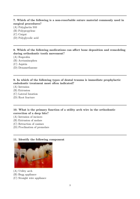
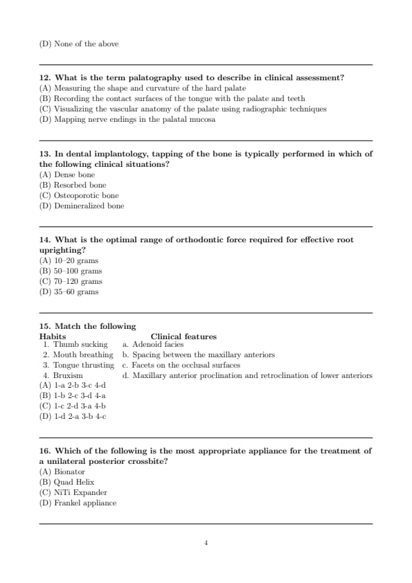
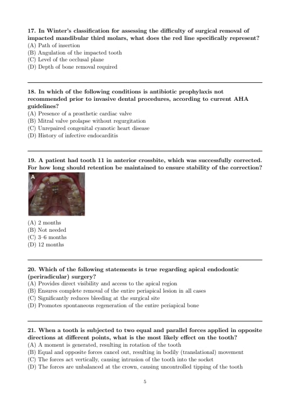
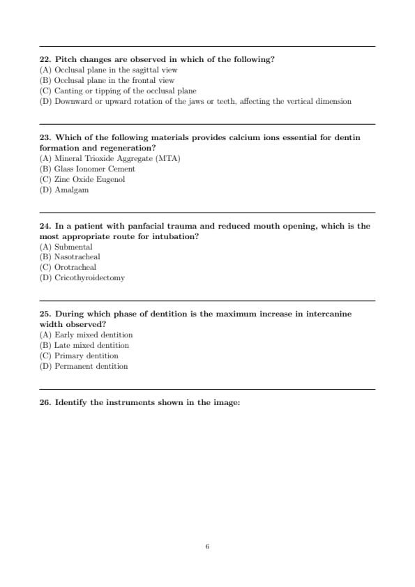
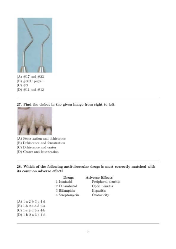
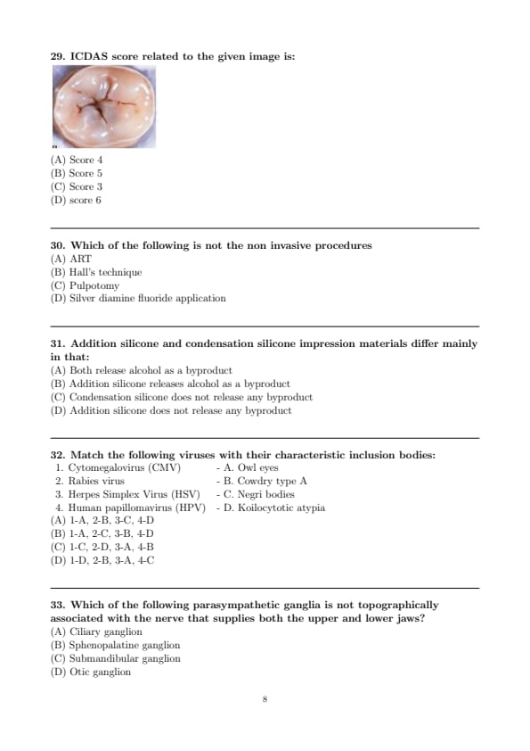
*The article might have information for the previous academic years, please refer the official website of the exam.