
NEET PG 2025 Question Paper with Solutions Pdf


Content Writer | Updated On - Mar 5, 2026
NEET PG 2025 Question Paper with Solutions is now available for download. The National Eligibility cum Entrance Test for Postgraduate (NEET PG), conducted by the National Testing Agency (NTA), serves as a national-level entrance examination for admission to MD, MS, and PG Diploma courses across India. The NEET PG 2025 examination was held in a single timed session with a total duration of 3 hours and 30 minutes, and the question paper comprised 200 multiple-choice questions, carrying a total of 800 marks. Candidates can now access the complete set of questions along with detailed solutions to help them evaluate their performance and understand the correct approach for each question.
NEET PG 2025 Question Paper with Solutions
| NEET PG 2025 Question Paper | Download PDF | Check Solutions |

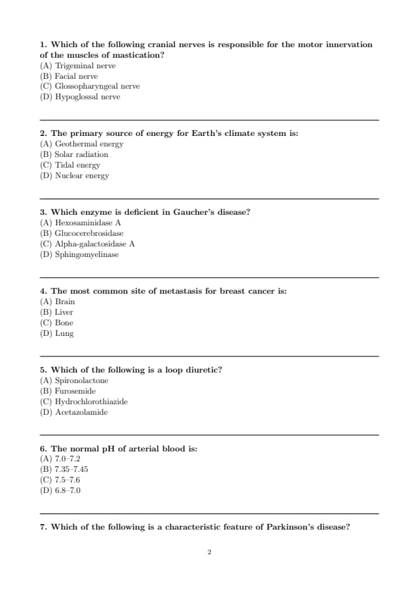
Which of the following cranial nerves is responsible for the motor innervation of the muscles of mastication?
View Solution
Step 1: Understanding the Concept:
The question asks to identify the cranial nerve that provides motor supply to the muscles responsible for chewing (mastication).
Step 3: Detailed Explanation:
The muscles of mastication include the masseter, temporalis, medial pterygoid, and lateral pterygoid. These muscles are responsible for the movements of the mandible at the temporomandibular joint, enabling chewing.
Let's analyze the functions of the listed cranial nerves:
Trigeminal nerve (CN V): This is a mixed nerve with both sensory and motor functions. Its motor component, specifically the mandibular division (V3), innervates the muscles of mastication. It also provides sensory information from the face and head.
Facial nerve (CN VII): This nerve is primarily responsible for the motor innervation of the muscles of facial expression. It also carries taste sensation from the anterior two-thirds of the tongue and provides parasympathetic innervation to salivary and lacrimal glands.
Glossopharyngeal nerve (CN IX): This nerve has mixed functions, including motor innervation to the stylopharyngeus muscle (which aids in swallowing), sensory information from the pharynx, and taste from the posterior one-third of the tongue.
Hypoglossal nerve (CN XII): This is a motor nerve that innervates the intrinsic and extrinsic muscles of the tongue, controlling tongue movements essential for speech and swallowing.
Based on this, the Trigeminal nerve (CN V) is the correct answer as it directly controls the muscles of mastication.
Step 4: Final Answer:
The cranial nerve responsible for the motor innervation of the muscles of mastication is the Trigeminal nerve.
Quick Tip: A useful mnemonic to remember the functions of cranial nerves is \textbf{"Some Say Marry Money But My Brother Says Big Brains Matter More"}. 'M' stands for motor, 'S' for sensory, and 'B' for both. The trigeminal nerve (CN V) is "Both", with its motor part supplying the muscles of mastication.
The primary source of energy for Earth's climate system is:
View Solution
Step 1: Understanding the Concept:
The question asks for the main energy source that drives the Earth's climate system, which includes atmospheric circulation, ocean currents, and the water cycle.
Step 3: Detailed Explanation:
Let's evaluate the energy sources listed:
Geothermal energy: This is heat originating from the Earth's core. While significant, its contribution to the surface temperature and climate system is very small compared to the energy received from the sun.
Solar radiation: This is the energy emitted by the sun that reaches Earth. It is the fundamental driver of Earth's weather and climate. Solar energy heats the Earth's surface unevenly, creating temperature differences that drive atmospheric winds and ocean currents. It also powers the water cycle through evaporation.
Tidal energy: This energy is derived from the gravitational pull of the moon and sun on Earth's oceans. It has localized effects but is not a primary driver of the global climate system.
Nuclear energy: This refers to the energy from radioactive decay within the Earth's interior, which is the source of geothermal heat. As mentioned, its impact on the climate system is minimal.
The vast majority of the energy in the Earth's atmosphere and oceans originates from the sun. Therefore, solar radiation is the primary source of energy for the climate system.
Step 4: Final Answer:
Solar radiation is the fundamental energy source that powers the Earth's climate system.
Quick Tip: Remember that almost all weather phenomena, from winds to rain, are driven by the differential heating of the Earth's surface by the sun. This makes solar radiation the cornerstone of climate science.
Which enzyme is deficient in Gaucher's disease?
View Solution
Step 1: Understanding the Concept:
The question is about identifying the specific enzyme deficiency that leads to Gaucher's disease, which is a type of lysosomal storage disorder.
Step 3: Detailed Explanation:
Lysosomal storage diseases are a group of genetic disorders caused by the deficiency of specific enzymes required for the breakdown of macromolecules in lysosomes. This deficiency leads to the accumulation of the substrate within cells, causing various clinical manifestations.
Let's analyze the options:
Hexosaminidase A: Deficiency of this enzyme causes Tay-Sachs disease, leading to the accumulation of GM2 ganglioside.
Glucocerebrosidase (or \(\beta\)-glucosidase): Deficiency of this enzyme causes Gaucher's disease. This results in the accumulation of glucocerebroside in cells of the mononuclear phagocyte system, leading to hepatosplenomegaly, bone disease, and anemia.
Alpha-galactosidase A: Deficiency of this enzyme causes Fabry disease, leading to the accumulation of globotriaosylceramide (Gb3).
Sphingomyelinase: Deficiency of this enzyme causes Niemann-Pick disease (Types A and B), resulting in the accumulation of sphingomyelin.
Therefore, the enzyme deficient in Gaucher's disease is Glucocerebrosidase.
Step 4: Final Answer:
Gaucher's disease is caused by a deficiency of the enzyme Glucocerebrosidase.
Quick Tip: For exams, create a table linking common lysosomal storage diseases with their deficient enzyme, accumulated substrate, and key clinical features. This helps in quick recall and differentiation.
The most common site of metastasis for breast cancer is:
View Solution
Step 1: Understanding the Concept:
The question asks to identify the most frequent site where breast cancer cells spread (metastasize) to from the primary tumor.
Step 3: Detailed Explanation:
Metastasis is the process by which cancer cells spread from the primary site to other parts of the body, typically through the bloodstream or lymphatic system. Breast cancer has a propensity to metastasize to specific distant organs.
The common sites for breast cancer metastasis, in order of frequency, are:
Bone: This is the most common site of distant metastasis for breast cancer. The spine, ribs, pelvis, and long bones are frequently affected. Bone metastases can be lytic (bone-destroying), blastic (bone-forming), or mixed.
Lung: The lungs are the second most common site.
Liver: The liver is another frequent site of metastasis.
Brain: Brain metastases are also common, particularly with certain subtypes of breast cancer like HER2-positive and triple-negative breast cancer.
While all the options listed are common sites for metastasis, bone is the most frequent.
Step 4: Final Answer:
The most common site of metastasis for breast cancer is the bone.
Quick Tip: Remember the common sites of breast cancer metastasis with the mnemonic "B.L.L.B." - \textbf{B}one, \textbf{L}ung, \textbf{L}iver, \textbf{B}rain. Bone is the most common destination for metastatic breast cancer cells.
Which of the following is a loop diuretic?
View Solution
Step 1: Understanding the Concept:
The question requires identifying which of the given drugs belongs to the class of loop diuretics, based on its mechanism and site of action in the kidney.
Step 3: Detailed Explanation:
Diuretics are drugs that increase urine output. They are classified based on their site of action within the nephron.
Spironolactone: This is a potassium-sparing diuretic. It acts as an aldosterone antagonist in the collecting ducts, promoting sodium and water excretion while retaining potassium.
Furosemide: This is a potent loop diuretic. It acts on the thick ascending limb of the Loop of Henle by inhibiting the Na-K-2Cl cotransporter. This action prevents the reabsorption of a significant amount of sodium, leading to a powerful diuretic effect. Other examples include bumetanide and torsemide.
Hydrochlorothiazide: This is a thiazide diuretic. It acts on the distal convoluted tubule by inhibiting the Na-Cl cotransporter, leading to a moderate diuretic effect.
Acetazolamide: This is a carbonic anhydrase inhibitor. It acts on the proximal convoluted tubule, causing weak diuresis by preventing bicarbonate reabsorption.
Among the options, Furosemide is the classic example of a loop diuretic.
Step 4: Final Answer:
Furosemide is a loop diuretic.
Quick Tip: To remember diuretic classes and their sites of action, visualize the nephron. Proximal tubule (Acetazolamide), Loop of Henle (Furosemide), Distal Convoluted Tubule (Hydrochlorothiazide), and Collecting Duct (Spironolactone).
The normal pH of arterial blood is:
View Solution
Step 1: Understanding the Concept:
This question tests the knowledge of a fundamental physiological parameter: the normal pH range of human arterial blood, which is critical for homeostasis.
Step 3: Detailed Explanation:
The pH is a measure of the acidity or alkalinity of a solution. The human body maintains the pH of blood in a very narrow and tightly regulated range to ensure optimal functioning of enzymes and metabolic processes.
The normal pH range for arterial blood is 7.35 to 7.45.
A pH below 7.35 is termed acidemia (indicating an underlying acidosis).
A pH above 7.45 is termed alkalemia (indicating an underlying alkalosis).
The ranges given in the other options are incompatible with life for any sustained period. For example, a pH below 6.8 or above 7.8 can be fatal.
Therefore, the correct range is 7.35–7.45.
Step 4: Final Answer:
The normal pH of arterial blood is between 7.35 and 7.45.
Quick Tip: This is a core value in medicine that must be memorized. For quick reference, remember the midpoint of 7.40. Any deviation from the 7.35-7.45 range indicates a significant acid-base disturbance.
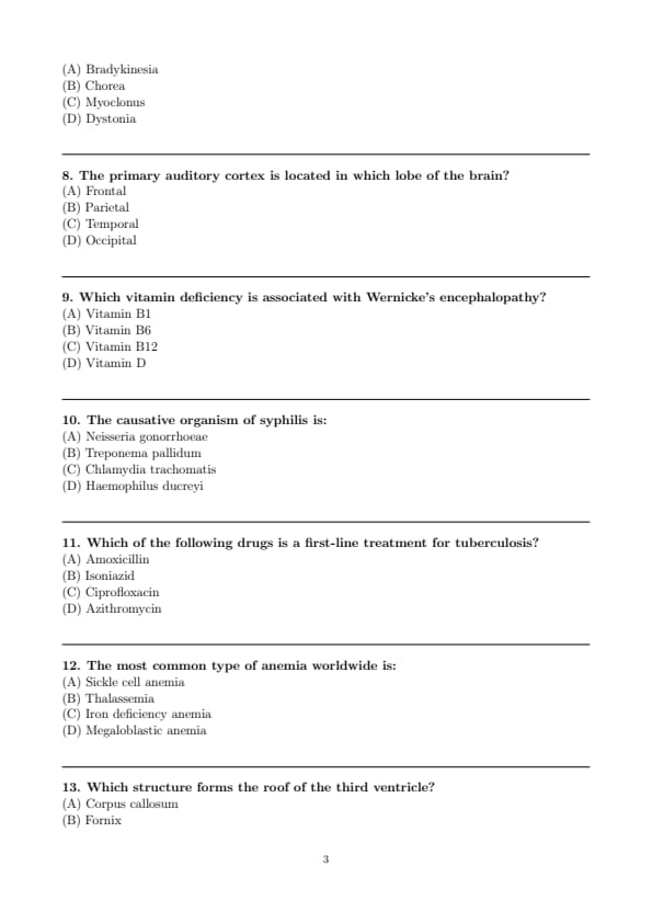
Which of the following is a characteristic feature of Parkinson's disease?
View Solution
Step 1: Understanding the Concept:
The question asks to identify a cardinal sign of Parkinson's disease from a list of movement disorders.
Step 3: Detailed Explanation:
Parkinson's disease is a neurodegenerative disorder primarily affecting dopaminergic neurons in the substantia nigra. It is characterized by a specific set of motor symptoms. Let's analyze the options:
Bradykinesia: This means "slowness of movement" and is a core, universal feature of Parkinson's disease. It manifests as difficulty initiating movements (akinesia) and a reduction in the speed and amplitude of voluntary movements.
Chorea: These are involuntary, rapid, irregular, and jerky movements. Chorea is the hallmark feature of Huntington's disease, not Parkinson's disease.
Myoclonus: These are sudden, brief, shock-like muscle jerks. They can be seen in various conditions, including epilepsy and Creutzfeldt-Jakob disease, but are not a primary feature of Parkinson's.
Dystonia: This involves sustained or intermittent muscle contractions causing abnormal, often repetitive, movements or postures. It can be a symptom in some Parkinson's patients but is not considered a cardinal feature like bradykinesia.
The four cardinal features of Parkinson's disease are summarized by the acronym TRAP: Tremor (at rest), Rigidity, Akinesia/Bradykinesia, and Postural instability. Bradykinesia is one of these defining characteristics.
Step 4: Final Answer:
Bradykinesia is a characteristic feature of Parkinson's disease.
Quick Tip: Use the mnemonic \textbf{TRAP} to remember the cardinal motor symptoms of Parkinson's disease: \textbf{T}remor, \textbf{R}igidity, \textbf{A}kinesia (or Bradykinesia), and \textbf{P}ostural instability. This will help you quickly identify the key features.
The primary auditory cortex is located in which lobe of the brain?
View Solution
Step 1: Understanding the Concept:
This question tests knowledge of functional neuroanatomy, specifically the location of the brain region responsible for processing primary auditory information.
Step 3: Detailed Explanation:
The cerebral cortex is divided into four main lobes, each with specialized functions.
Frontal Lobe: Associated with higher executive functions, planning, motor control (primary motor cortex), and speech production (Broca's area).
Parietal Lobe: Responsible for processing somatosensory information (touch, temperature, pain) in the primary somatosensory cortex, spatial awareness, and navigation.
Temporal Lobe: This lobe is crucial for auditory processing, memory, and language comprehension. The primary auditory cortex (Brodmann areas 41 and 42) is located in the superior temporal gyrus of this lobe. It is the first cortical region to receive and process auditory information from the ears. Wernicke's area, critical for language comprehension, is also located here.
Occipital Lobe: This is the primary center for visual processing, containing the primary visual cortex.
Therefore, the primary auditory cortex is located in the temporal lobe.
Step 4: Final Answer:
The primary auditory cortex is located in the temporal lobe of the brain.
Quick Tip: A simple way to remember the primary sensory areas: Occipital lobe is for sight (think of "optical"), Temporal lobe is for hearing (near the temples/ears), and Parietal lobe is for touch/sensation.
Which vitamin deficiency is associated with Wernicke's encephalopathy?
View Solution
Step 1: Understanding the Concept:
The question asks to identify the specific vitamin deficiency that causes the neurological disorder known as Wernicke's encephalopathy.
Step 3: Detailed Explanation:
Wernicke's encephalopathy is an acute and life-threatening neurological condition. Let's examine the roles of the vitamins listed:
Vitamin B1 (Thiamine): Thiamine is a crucial coenzyme in glucose metabolism in the brain. Its deficiency impairs cerebral energy utilization, leading to neuronal damage. This deficiency is the direct cause of Wernicke's encephalopathy, classically presenting with a triad of symptoms: confusion, ophthalmoplegia (abnormal eye movements), and ataxia (gait disturbance). It is most commonly seen in individuals with chronic alcoholism and malnutrition.
Vitamin B6 (Pyridoxine): Deficiency can cause peripheral neuropathy, seizures, and sideroblastic anemia.
Vitamin B12 (Cobalamin): Deficiency leads to megaloblastic anemia and subacute combined degeneration of the spinal cord, causing neurological symptoms like paresthesias and gait problems.
Vitamin D: Deficiency primarily causes bone disorders like rickets in children and osteomalacia in adults.
The condition specifically linked to Vitamin B1 deficiency is Wernicke's encephalopathy. If left untreated, it can progress to Korsakoff syndrome, a chronic memory disorder.
Step 4: Final Answer:
Wernicke's encephalopathy is associated with a deficiency of Vitamin B1 (Thiamine).
Quick Tip: Remember the classic triad of Wernicke's encephalopathy: \textbf{Confusion, Ophthalmoplegia, and Ataxia (COA)}. Associate this triad with Vitamin B1 (Thiamine) deficiency, especially in the context of alcoholism.
The causative organism of syphilis is:
View Solution
Step 1: Understanding the Concept:
The question requires the identification of the specific bacterium that causes the sexually transmitted infection (STI) syphilis.
Step 3: Detailed Explanation:
Let's identify the diseases caused by each of the organisms listed:
Neisseria gonorrhoeae: This is a gram-negative diplococcus that causes gonorrhea, another common STI.
Treponema pallidum: This is a spirochete (spiral-shaped) bacterium that is the causative agent of syphilis. Syphilis is a multi-stage disease that can affect various organs if left untreated.
Chlamydia trachomatis: This is an obligate intracellular bacterium that causes chlamydia, one of the most common STIs worldwide. It can also cause trachoma (an eye infection).
Haemophilus ducreyi: This is a gram-negative coccobacillus that causes chancroid, an STI characterized by painful genital ulcers.
Thus, the causative organism of syphilis is Treponema pallidum.
Step 4: Final Answer:
The bacterium that causes syphilis is \textit{Treponema pallidum.
Quick Tip: For STIs, it's crucial to link the disease to its causative organism and key clinical feature. For example: Syphilis -> \textit{T. pallidum -> painless chancre; Chancroid -> H. ducreyi -> painful ulcer.
Which of the following drugs is a first-line treatment for tuberculosis?
View Solution
Step 1: Understanding the Concept:
The question asks to identify a drug that is part of the standard, first-line regimen for treating active tuberculosis (TB).
Step 3: Detailed Explanation:
The treatment of active tuberculosis requires a multi-drug regimen to prevent the development of drug resistance. The standard first-line therapy consists of four drugs. Let's analyze the options:
Amoxicillin: This is a beta-lactam antibiotic, commonly used for various bacterial infections, but it is not effective against Mycobacterium tuberculosis.
Isoniazid (INH): This is a cornerstone of TB treatment. It is a bactericidal agent that inhibits the synthesis of mycolic acids, which are essential components of the mycobacterial cell wall. It is a key component of the first-line regimen.
Ciprofloxacin: This is a fluoroquinolone antibiotic. Fluoroquinolones are considered second-line agents for TB, used in cases of resistance to first-line drugs or intolerance.
Azithromycin: This is a macrolide antibiotic, used for various respiratory and other infections, including infections caused by atypical mycobacteria, but it is not a first-line drug for \textit{M. tuberculosis.
The standard first-line regimen for active TB is known as RIPE: Rifampicin, Isoniazid, Pyrazinamide, and Ethambutol. Isoniazid is a critical part of this combination.
Step 4: Final Answer:
Isoniazid is a first-line drug for the treatment of tuberculosis.
Quick Tip: Memorize the acronym \textbf{RIPE for the four first-line anti-tuberculosis drugs: \textbf{R}ifampicin, \textbf{I}soniazid, \textbf{P}yrazinamide, and \textbf{E}thambutol. Also, know their major side effects (e.g., Isoniazid - neuropathy, hepatotoxicity; Rifampicin - orange body fluids, hepatotoxicity).
The most common type of anemia worldwide is:
View Solution
Step 1: Understanding the Concept:
The question asks to identify the most prevalent form of anemia on a global scale.
Step 3: Detailed Explanation:
Anemia is a condition characterized by a reduced number of red blood cells or a lower-than-normal quantity of hemoglobin. Let's consider the options:
Sickle cell anemia: A genetic hemoglobinopathy common in populations of African, Mediterranean, and South Asian descent, but not the most common worldwide.
Thalassemia: A group of genetic blood disorders affecting hemoglobin production, prevalent in similar regions to sickle cell disease, but less common overall than iron deficiency.
Iron deficiency anemia: This is the most common nutritional disorder in the world and the leading cause of anemia globally. It is caused by insufficient dietary intake of iron, poor absorption, or chronic blood loss (e.g., from menstruation or gastrointestinal bleeding). It affects a vast number of people, especially women and children in developing countries.
Megaloblastic anemia: Caused by a deficiency of Vitamin B12 or folate, leading to impaired DNA synthesis and large, immature red blood cells. While significant, its prevalence is lower than that of iron deficiency anemia.
Due to its strong link with nutrition and chronic blood loss, iron deficiency anemia is by far the most common type of anemia worldwide.
Step 4: Final Answer:
The most common type of anemia worldwide is iron deficiency anemia.
Quick Tip: When thinking about global health statistics, always consider nutritional deficiencies first for conditions like anemia. Iron deficiency affects billions of people, making it the most common cause.
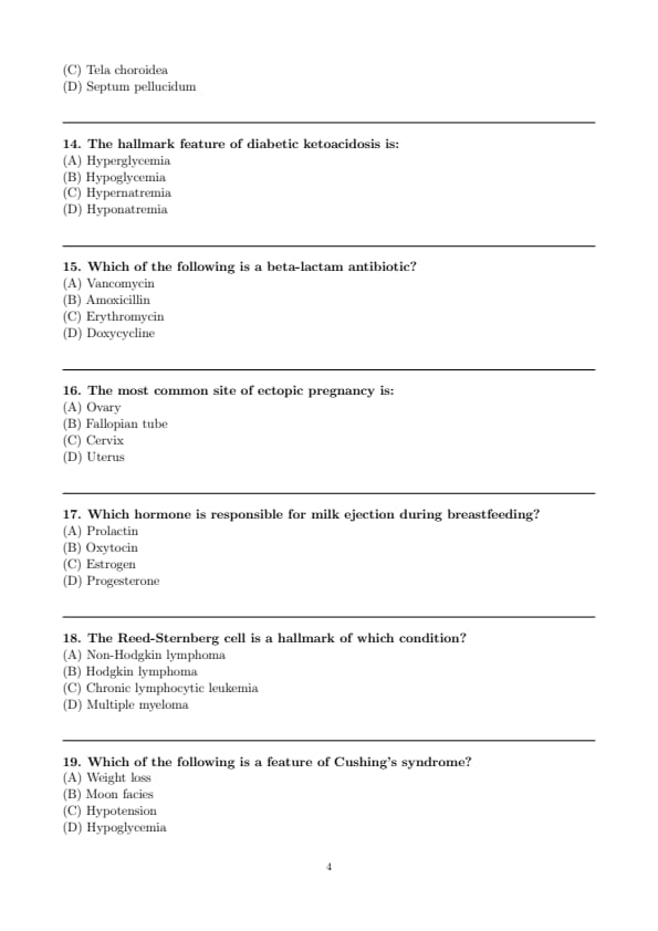
Which structure forms the roof of the third ventricle?
View Solution
Step 1: Understanding the Concept:
This question tests detailed knowledge of the anatomy of the ventricular system of the brain, specifically the structures that form the boundaries of the third ventricle.
Step 3: Detailed Explanation:
The third ventricle is a narrow, midline cavity in the diencephalon, filled with cerebrospinal fluid. Its boundaries are formed by various important brain structures. Let's analyze the structures listed in relation to the roof:
Corpus callosum: This is a large white matter tract that connects the two cerebral hemispheres. It lies superior to the fornix and the third ventricle, but does not form the direct roof.
Fornix: This is a C-shaped bundle of nerve fibers that is a major output tract of the hippocampus. The body of the fornix runs superior to the thalamus and forms the superior boundary or landmark for the roof of the third ventricle, but it is not the roof itself.
Tela choroidea: This is a thin membrane of pia mater and ependyma that forms the true, immediate roof of the third ventricle. The choroid plexus of the third ventricle, which produces CSF, hangs down from this membrane into the ventricle.
Septum pellucidum: This is a thin vertical membrane that separates the anterior horns of the left and right lateral ventricles. It does not form the roof of the third ventricle.
Therefore, the direct roof of the third ventricle is the tela choroidea, with the fornix and corpus callosum located superior to it.
Step 4: Final Answer:
The structure that forms the roof of the third ventricle is the tela choroidea.
Quick Tip: When studying neuroanatomy, use sagittal and coronal diagrams to visualize the relationships between deep brain structures. For the third ventricle, remember its roof is a delicate membrane (tela choroidea) from which the choroid plexus arises, located just beneath the fornix.
The hallmark feature of diabetic ketoacidosis is:
View Solution
Step 1: Understanding the Concept:
The question asks for a defining characteristic of diabetic ketoacidosis (DKA), a serious acute complication of diabetes mellitus.
Step 3: Detailed Explanation:
Diabetic ketoacidosis (DKA) is a state of absolute or relative insulin deficiency. This deficiency leads to a specific triad of metabolic disturbances that define the condition:
Hyperglycemia: Without sufficient insulin, glucose cannot enter cells for energy, so it accumulates in the bloodstream. Blood glucose levels are typically very high (e.g., > 250 mg/dL).
Ketosis: The body, starved for glucose, begins to break down fat for energy. This process produces acidic ketone bodies (e.g., acetoacetate, \(\beta\)-hydroxybutyrate), leading to ketonemia and ketonuria.
Metabolic Acidosis: The accumulation of acidic ketone bodies overwhelms the body's buffering capacity, causing the blood pH to drop (typically < 7.3).
Let's look at the options:
(A) Hyperglycemia: This is a core and initiating feature of DKA.
(B) Hypoglycemia: This is low blood sugar, the opposite of what occurs in DKA. It is more associated with insulin overdose.
(C) Hypernatremia & (D) Hyponatremia: Sodium levels can be variable in DKA. Osmotic diuresis from hyperglycemia can lead to total body sodium and water loss, potentially causing hypernatremia. However, severe hyperglycemia can also cause a dilutional or "pseudohyponatremia". While electrolyte abnormalities are present, they are not the primary hallmark feature like hyperglycemia.
Therefore, hyperglycemia is a fundamental hallmark of DKA.
Step 4: Final Answer:
The hallmark feature of diabetic ketoacidosis is hyperglycemia.
Quick Tip: Remember the diagnostic triad for DKA: \textbf{Hyperglycemia}, \textbf{Ketosis}, and \textbf{Acidosis}. The "D" in DKA can remind you of "Diabetic," which implies high blood sugar.
Which of the following is a beta-lactam antibiotic?
View Solution
Step 1: Understanding the Concept:
The question requires the classification of antibiotics, specifically identifying which of the given drugs belongs to the beta-lactam class.
Step 3: Detailed Explanation:
Beta-lactam antibiotics are a broad class of drugs that share a common chemical structure: the beta-lactam ring. They work by inhibiting bacterial cell wall synthesis. This class includes several major groups. Let's examine the options:
Vancomycin: This is a glycopeptide antibiotic. It also inhibits cell wall synthesis but through a different mechanism than beta-lactams (it binds to the D-Ala-D-Ala terminus of peptidoglycan precursors).
Amoxicillin: This is a member of the penicillin family (specifically an aminopenicillin). Penicillins are the original and most well-known group of beta-lactam antibiotics. Therefore, Amoxicillin is a beta-lactam antibiotic.
Erythromycin: This is a macrolide antibiotic. Macrolides work by inhibiting bacterial protein synthesis by binding to the 50S ribosomal subunit. They do not have a beta-lactam ring.
Based on this classification, Amoxicillin is the beta-lactam antibiotic among the choices.
Step 4: Final Answer:
Amoxicillin is a beta-lactam antibiotic.
Quick Tip: Memorize the major antibiotic classes and one or two key examples for each. The main Beta-Lactam groups are Penicillins (Amoxicillin, Piperacillin), Cephalosporins (Ceftriaxone), Carbapenems (Imipenem), and Monobactams (Aztreonam).
The most common site of ectopic pregnancy is:
View Solution
Step 1: Understanding the Concept:
An ectopic pregnancy is a pregnancy in which the fertilized egg implants outside the main cavity of the uterus. The question asks for the most frequent location for this abnormal implantation.
Step 3: Detailed Explanation:
Normally, a fertilized egg travels down the fallopian tube and implants in the uterine wall. In an ectopic pregnancy, this process is disrupted. Let's look at the possible sites:
Uterus: Implantation within the uterus is a normal pregnancy, not ectopic. (Note: A cornual/interstitial pregnancy is a rare type of uterine ectopic pregnancy, but the main uterine cavity is the normal site).
Fallopian tube: This is overwhelmingly the most common site for an ectopic pregnancy, accounting for over 95% of all cases. The fertilized egg gets stuck in the tube, often due to damage or blockage. The most common location within the tube is the ampulla.
Ovary: Ovarian pregnancies are rare, accounting for about 1-3% of ectopic pregnancies.
Cervix: Cervical pregnancies are also very rare (less than 1%).
Other rare sites include the abdominal cavity and cesarean scar. Given the statistics, the fallopian tube is the most common site by a large margin.
Step 4: Final Answer:
The most common site of an ectopic pregnancy is the fallopian tube.
Quick Tip: For exams, "ectopic pregnancy" should be almost synonymous with "tubal pregnancy" because it is so common. Risk factors that damage the fallopian tubes, like Pelvic Inflammatory Disease (PID) or previous tubal surgery, increase the risk of ectopic pregnancy.
Which hormone is responsible for milk ejection during breastfeeding?
View Solution
Step 1: Understanding the Concept:
The question asks to identify the specific hormone that causes the "let-down" reflex, which is the ejection of milk from the mammary glands during breastfeeding.
Step 3: Detailed Explanation:
Lactation is controlled by two key hormones with distinct roles:
Prolactin: Secreted by the anterior pituitary gland, prolactin is the primary hormone responsible for milk production (lactogenesis). Its levels rise after delivery and are stimulated by suckling. So, Prolactin = Production.
Oxytocin: Secreted by the posterior pituitary gland, oxytocin is responsible for milk ejection. The physical stimulus of suckling on the nipple sends signals to the hypothalamus, which triggers the release of oxytocin. Oxytocin then travels to the breast and causes the contraction of myoepithelial cells surrounding the alveoli, squeezing the milk out into the ducts. So, Oxytocin = Ejection.
Estrogen and Progesterone: During pregnancy, high levels of these hormones stimulate the development of the mammary glands but actually inhibit the action of prolactin on milk production. Their levels drop sharply after delivery, allowing lactation to begin.
Therefore, oxytocin is the hormone responsible for the milk ejection reflex.
Step 4: Final Answer:
Oxytocin is responsible for milk ejection during breastfeeding.
Quick Tip: A simple mnemonic to differentiate the roles is: \textbf{P}rolactin is for \textbf{P}roduction, while \textbf{O}xytocin makes the milk flow \textbf{O}ut.
The Reed-Sternberg cell is a hallmark of which condition?
View Solution
N/A Quick Tip: Associate the "owl's eye" appearance with the Reed-Sternberg cell, and remember that this cell is the defining feature that separates Hodgkin lymphoma from the much broader category of Non-Hodgkin lymphomas.
Which of the following is a feature of Cushing's syndrome?
View Solution
Step 1: Understanding the Concept:
The question asks to identify a clinical sign characteristic of Cushing's syndrome, a condition caused by prolonged exposure to high levels of cortisol.
Step 3: Detailed Explanation:
Cushing's syndrome results from excess glucocorticoids (cortisol). The widespread effects of cortisol on metabolism and body composition lead to a constellation of characteristic signs and symptoms. Let's analyze the options:
Weight loss: This is incorrect. Cortisol promotes fat deposition, leading to central obesity (truncal obesity) and overall weight gain.
Moon facies: This is a classic feature of Cushing's syndrome. It describes a round, full, and red face due to the redistribution of fat to the face and cheeks.
Hypotension: This is incorrect. Cortisol increases vascular sensitivity to catecholamines and has mineralocorticoid effects, leading to hypertension.
Hypoglycemia: This is incorrect. Cortisol is a glucocorticoid that promotes gluconeogenesis and insulin resistance, leading to hyperglycemia.
Other classic features include a "buffalo hump" (fat pad on the upper back), purple striae on the abdomen, muscle weakness, and easy bruising. "Moon facies" is one of the most recognizable signs.
Step 4: Final Answer:
Moon facies is a feature of Cushing's syndrome.
Quick Tip: Remember the key features of Cushing's syndrome by thinking of the "Cushingoid" appearance: \textbf{C}entral obesity, \textbf{U}ncontrolled blood sugar and pressure, \textbf{S}triae, \textbf{H}irsutism, \textbf{I}mmunosuppression, \textbf{N}eck fat pad (buffalo hump), and \textbf{G}rowth retardation, along with Moon facies.
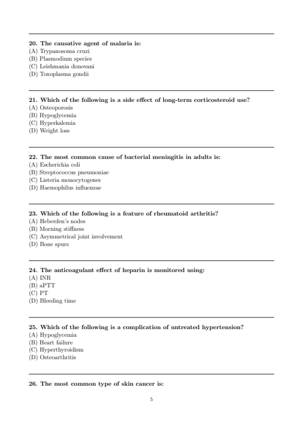
The causative agent of malaria is:
View Solution
Step 1: Understanding the Concept:
This question asks for the specific pathogen responsible for causing the infectious disease malaria.
Step 3: Detailed Explanation:
Malaria is a mosquito-borne parasitic disease. Let's identify the diseases caused by the listed organisms:
Trypanosoma cruzi: This is a protozoan parasite that causes Chagas disease (American trypanosomiasis), transmitted by triatomine bugs ("kissing bugs").
Plasmodium species: These are protozoan parasites that cause malaria. The disease is transmitted by the bite of infected female Anopheles mosquitoes. The five species that cause malaria in humans are \textit{P. falciparum, \textit{P. vivax, \textit{P. ovale, \textit{P. malariae, and \textit{P. knowlesi. \textit{P. falciparum is the most deadly.
Leishmania donovani: This protozoan parasite causes visceral leishmaniasis (kala-azar), transmitted by the bite of infected sandflies.
Toxoplasma gondii: This protozoan parasite causes toxoplasmosis, which is commonly acquired through ingestion of contaminated food or water, or contact with infected cat feces.
Therefore, the causative agent of malaria is the \textit{Plasmodium species.
Step 4: Final Answer:
The causative agent of malaria is the Plasmodium species.
Quick Tip: It is essential to link major parasitic diseases with their causative genus. For your exam, remember: \textit{Plasmodium = Malaria, Trypanosoma = Chagas/Sleeping Sickness, Leishmania = Leishmaniasis, Toxoplasma = Toxoplasmosis.
Which of the following is a side effect of long-term corticosteroid use?
View Solution
Step 1: Understanding the Concept:
The question asks to identify a known adverse effect resulting from the chronic administration of corticosteroid medications (e.g., prednisone, dexamethasone).
Step 3: Detailed Explanation:
Long-term corticosteroid use leads to numerous and significant side effects due to their widespread physiological actions. This is often referred to as iatrogenic Cushing's syndrome. Let's analyze the options:
Osteoporosis: This is a major and common side effect. Corticosteroids decrease bone formation by inhibiting osteoblast function and increase bone resorption by promoting osteoclast activity. They also reduce intestinal calcium absorption. This combination leads to a loss of bone density and an increased risk of fractures.
Hypoglycemia (low blood sugar): This is incorrect. Corticosteroids are glucocorticoids, meaning they raise blood glucose levels by promoting gluconeogenesis and causing insulin resistance. Thus, hyperglycemia is the side effect.
Hyperkalemia (high potassium): This is incorrect. Corticosteroids, particularly those with mineralocorticoid activity, can promote potassium excretion by the kidneys, leading to hypokalemia.
Weight loss: This is incorrect. Corticosteroids stimulate appetite and cause fat redistribution (central obesity), leading to weight gain.
Therefore, osteoporosis is a well-established side effect of long-term corticosteroid therapy.
Step 4: Final Answer:
Osteoporosis is a side effect of long-term corticosteroid use.
Quick Tip: Remember that corticosteroids have effects on "sugar, salt, and sex" (glucocorticoid, mineralocorticoid, and androgenic effects). They increase blood sugar (hyperglycemia), can cause low potassium (hypokalemia), and lead to bone loss (osteoporosis).
The most common cause of bacterial meningitis in adults is:
View Solution
Step 1: Understanding the Concept:
The question asks to identify the most frequent bacterial pathogen causing meningitis in the adult population.
Step 3: Detailed Explanation:
The epidemiology of bacterial meningitis varies significantly with age. Let's examine the common causative agents:
Escherichia coli: Along with Group B Streptococcus, this is a leading cause of meningitis in neonates, often acquired during birth.
Streptococcus pneumoniae (Pneumococcus): This is the most common cause of community-acquired bacterial meningitis in adults and the elderly. It is also a common cause in children over the age of 2.
Listeria monocytogenes: This pathogen typically causes meningitis in individuals at the extremes of age (neonates and the elderly) and those who are immunocompromised.
Haemophilus influenzae type b (Hib): This was historically a major cause of meningitis in young children, but its incidence has dramatically decreased in many countries due to the widespread use of the Hib vaccine.
In a typical adult patient, Streptococcus pneumoniae is the most likely causative organism, followed by \textit{Neisseria meningitidis (which is common in adolescents and young adults, especially in epidemic settings like dormitories).
Step 4: Final Answer:
The most common cause of bacterial meningitis in adults is \textit{Streptococcus pneumoniae.
Quick Tip: Remember the common meningitis bugs by age group: \textbf{Neonates: Group B Strep, E. coli, Listeria. \textbf{Children/Young Adults:} Neisseria meningitidis, S. pneumoniae. \textbf{Adults/Elderly:} S. pneumoniae, N. meningitidis, Listeria.
Which of the following is a feature of rheumatoid arthritis?
View Solution
Step 1: Understanding the Concept:
The question asks to identify a clinical feature that is characteristic of rheumatoid arthritis (RA), an autoimmune inflammatory disease, and distinguish it from features of other arthritides like osteoarthritis (OA).
Step 3: Detailed Explanation:
Rheumatoid arthritis is a systemic inflammatory disorder that primarily affects synovial joints. Let's analyze the options:
Heberden's nodes: These are bony swellings (osteophytes) of the distal interphalangeal (DIP) joints. They are a classic sign of osteoarthritis, not RA. (Nodes at the proximal interphalangeal joints are called Bouchard's nodes, also seen in OA).
Morning stiffness: This is a hallmark feature of inflammatory arthritis like RA. The stiffness is typically prolonged, lasting for more than one hour upon waking. This is due to the accumulation of inflammatory fluid in the joints overnight. In contrast, the morning stiffness in OA is usually brief (less than 30 minutes).
Asymmetrical joint involvement: This is more characteristic of osteoarthritis. RA classically presents with symmetrical involvement, meaning if a joint on one side of the body is affected, the same joint on the other side is also likely to be affected.
Bone spurs (osteophytes): These are bony projections that form along joint margins. They are a defining feature of the degenerative process in osteoarthritis, not RA. RA is characterized by erosions, not osteophytes.
Therefore, prolonged morning stiffness is a key feature of rheumatoid arthritis.
Step 4: Final Answer:
Morning stiffness is a feature of rheumatoid arthritis.
Quick Tip: Create a comparison table for RA vs. OA. Key differentiating points: \textbf{RA:} Autoimmune, Symmetrical, Inflammatory, Morning stiffness >1hr, Erosions. \textbf{OA:} Degenerative, Asymmetrical, Non-inflammatory, Morning stiffness <30min, Osteophytes (bone spurs).
The anticoagulant effect of heparin is monitored using:
View Solution
Step 1: Understanding the Concept:
The question asks for the specific laboratory test used to monitor the therapeutic level of unfractionated heparin, a commonly used anticoagulant.
Step 3: Detailed Explanation:
Different anticoagulants affect different parts of the coagulation cascade and thus require different monitoring tests.
Heparin (unfractionated): It works by potentiating the action of antithrombin III, which then inactivates several clotting factors, most notably thrombin (Factor IIa) and Factor Xa. Its primary effect is on the intrinsic pathway of the coagulation cascade. The test that measures the integrity of the intrinsic pathway is the activated Partial Thromboplastin Time (aPTT). Therefore, aPTT is used to monitor unfractionated heparin therapy.
Prothrombin Time (PT) and International Normalized Ratio (INR): These tests measure the integrity of the extrinsic pathway. They are used to monitor the effect of warfarin, an oral anticoagulant that inhibits vitamin K-dependent clotting factors (II, VII, IX, X).
Bleeding time: This test assesses platelet function and primary hemostasis. It is not used to monitor heparin or warfarin.
Note: Low-molecular-weight heparin (LMWH) does not typically require monitoring, but if needed, an anti-Factor Xa assay is used.
Step 4: Final Answer:
The anticoagulant effect of heparin is monitored using the aPTT.
Quick Tip: A useful mnemonic: \textbf{WEPT} (\textbf{W}arfarin affects \textbf{E}xtrinsic pathway, monitor with \textbf{PT}). For heparin, remember it affects the intrinsic pathway, so you monitor it with the other test, aPTT.
Which of the following is a complication of untreated hypertension?
View Solution
Step 1: Understanding the Concept:
The question asks to identify a long-term consequence or complication that can arise from chronically elevated blood pressure (hypertension) that is not managed.
Step 3: Detailed Explanation:
Untreated hypertension leads to progressive damage to blood vessels and vital organs throughout the body. This is known as end-organ damage. Let's analyze the options:
Hypoglycemia: This is low blood sugar and is not related to hypertension. It's typically a concern in diabetes management.
Heart failure: This is a major complication of chronic hypertension. High blood pressure increases the afterload, which is the resistance the heart must pump against. Over time, this forces the left ventricle to work harder, leading to left ventricular hypertrophy (thickening of the heart muscle). Eventually, the hypertrophied muscle can become stiff (diastolic dysfunction) and/or weaken (systolic dysfunction), leading to heart failure.
Hyperthyroidism: This is a condition of excess thyroid hormone production and is not a complication of hypertension.
Osteoarthritis: This is a degenerative joint disease and is not caused by high blood pressure.
Other major complications of untreated hypertension include myocardial infarction (heart attack), stroke, chronic kidney disease, and hypertensive retinopathy.
Step 4: Final Answer:
Heart failure is a major complication of untreated hypertension.
Quick Tip: Remember the main target organs of hypertensive damage: the \textbf{Heart} (heart failure, MI), \textbf{Brain} (stroke), \textbf{Kidneys} (chronic kidney disease), and \textbf{Eyes} (retinopathy).
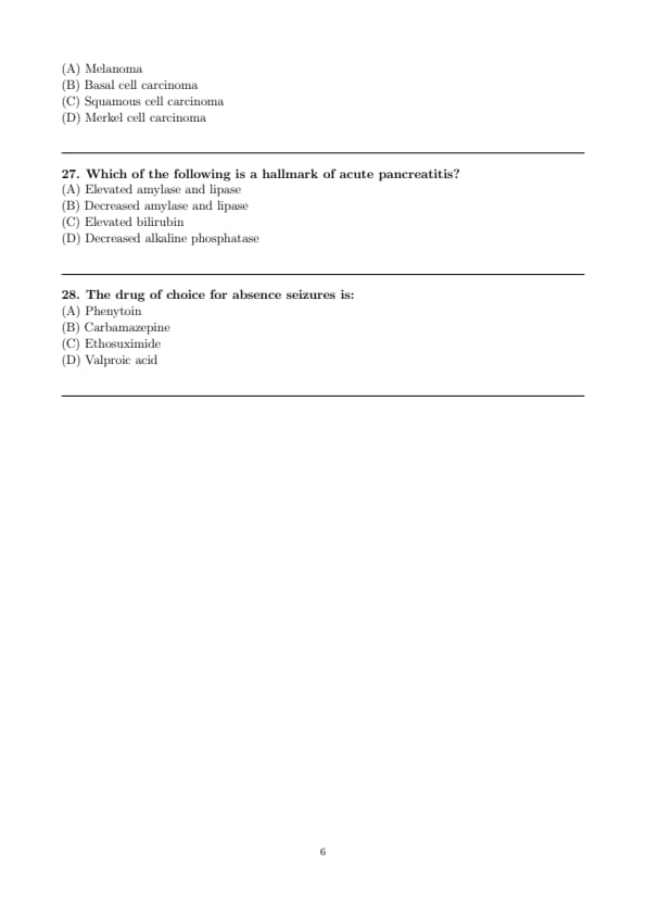
The most common type of skin cancer is:
View Solution
Step 1: Understanding the Concept:
The question asks to identify the most frequently diagnosed type of skin cancer among the given options.
Step 3: Detailed Explanation:
Skin cancers are broadly categorized into melanoma and non-melanoma skin cancers. Let's look at the prevalence of each type:
Basal cell carcinoma (BCC): This is the most common type of non-melanoma skin cancer and the most common cancer in humans overall. It arises from the basal cells in the epidermis. BCCs are typically slow-growing, locally invasive, and have a very low potential for metastasis.
Squamous cell carcinoma (SCC): This is the second most common type of skin cancer. It arises from squamous cells in the epidermis. SCCs have a higher risk of metastasis than BCCs, but the risk is still relatively low.
Melanoma: This cancer arises from melanocytes, the pigment-producing cells. While it is much less common than BCC and SCC, it is the most dangerous type of skin cancer because it has a high propensity to metastasize.
Merkel cell carcinoma: This is a rare but highly aggressive neuroendocrine cancer of the skin with a high risk of recurrence and metastasis.
Based on incidence, Basal cell carcinoma is by far the most common.
Step 4: Final Answer:
The most common type of skin cancer is Basal cell carcinoma.
Quick Tip: Remember the order of frequency for skin cancers: \textbf{Basal Cell > Squamous Cell > Melanoma}. However, the order of mortality is the reverse: \textbf{Melanoma > Squamous Cell > Basal Cell}.
Which of the following is a hallmark of acute pancreatitis?
View Solution
Step 1: Understanding the Concept:
The question asks for a key diagnostic finding in acute pancreatitis, an inflammatory condition of the pancreas.
Step 3: Detailed Explanation:
The diagnosis of acute pancreatitis is typically based on meeting at least two of the following three criteria:
Characteristic abdominal pain (acute onset, severe, epigastric pain often radiating to the back).
Biochemical evidence of pancreatitis, which is a significant elevation in serum amylase and/or lipase levels.
Characteristic findings on imaging (e.g., CT, MRI, ultrasound).
Let's evaluate the options:
(A) Elevated amylase and lipase: This is the key biochemical hallmark. In acute pancreatitis, inflammation and damage to pancreatic acinar cells lead to the release of these digestive enzymes into the bloodstream. A level of three times the upper limit of normal is generally considered diagnostic. Lipase is more specific for pancreatitis than amylase.
(B) Decreased amylase and lipase: This is incorrect. The levels are elevated.
(C) Elevated bilirubin: This can occur if the pancreatitis is caused by a gallstone obstructing the common bile duct (gallstone pancreatitis), leading to cholestasis. However, it is not a universal or primary hallmark of all cases of pancreatitis.
(D) Decreased alkaline phosphatase: This is incorrect. Alkaline phosphatase may be elevated in cases of biliary obstruction but would not be decreased.
Therefore, the most direct and common hallmark is the elevation of pancreatic enzymes.
Step 4: Final Answer:
Elevated amylase and lipase is a hallmark of acute pancreatitis.
Quick Tip: For acute pancreatitis, remember the two key diagnostic elements: severe epigastric pain radiating to the back and a serum lipase level greater than three times the upper limit of normal.
The drug of choice for absence seizures is:
View Solution
Step 1: Understanding the Concept:
The question asks to identify the first-line medication for treating a specific type of seizure known as an absence seizure (formerly petit mal seizure).
Step 3: Detailed Explanation:
Absence seizures are a type of generalized seizure characterized by brief episodes of staring and loss of awareness, without loss of postural tone. The choice of anti-epileptic drug is highly dependent on the seizure type.
Phenytoin and Carbamazepine: These are sodium channel blockers. They are effective for focal seizures and generalized tonic-clonic seizures, but they are not effective for absence seizures and can even worsen them.
Ethosuximide: This drug works by blocking T-type calcium channels in the thalamus, which are thought to be critical in the generation of the 3-Hz spike-and-wave discharges seen on EEG during absence seizures. It is highly specific and effective for absence seizures and is considered the first-line drug of choice for uncomplicated absence seizures, especially in children, due to its favorable side-effect profile compared to valproic acid.
Valproic acid (Valproate): This is a broad-spectrum anti-epileptic drug that is also effective against absence seizures (it also blocks T-type calcium channels, among other mechanisms). It is a first-line option and is preferred if the patient has both absence and generalized tonic-clonic seizures. However, for pure absence seizures, ethosuximide is often preferred.
Given the options, Ethosuximide is the classic and most specific drug of choice for absence seizures.
Step 4: Final Answer:
The drug of choice for absence seizures is Ethosuximide.
Quick Tip: For seizure types, remember: \textbf{Ethosuximide} is for pure \textbf{E}mpty spells (absence seizures). Be aware that some drugs, like Carbamazepine and Phenytoin, can paradoxically worsen this seizure type.

*The article might have information for the previous academic years, please refer the official website of the exam.