
NEET PG 2026 Question Paper with Solution PDF


Content Curator | Updated On - Sep 1, 2026
NEET PG 2026 Question Paper with Solutions will be available for download. The National Eligibility cum Entrance Test for Postgraduate (NEET PG), conducted by the NBEMS, serves as a national-level entrance examination for admission to MD, MS, and PG Diploma courses across India. The NEET PG 2026 examination will be held on August 30, from 9 am to 12:30 pm in CBT mode. The total duration of the exam is 3 hours and 30 minutes, and the question paper comprises of 180 multiple-choice questions, carrying a total of 720 marks.
NEET PG 2026 Question Paper with Solutions
| NEET PG 2026 Question Paper | Download PDF | Check Solutions |
Atomic Energy Regulatory Board (AERB) clearance or registration is NOT required for which of the following diagnostic modalities?
View Solution
Step 1: Understanding the Question:
The question asks to identify which medical imaging modality does not require registration or clearance from the Atomic Energy Regulatory Board (AERB) of India. The AERB is the national regulatory body that oversees the safety and use of ionizing radiation.
Step 2: Key Formula or Approach:
The AERB's mandate is restricted to facilities and equipment that utilize ionizing radiation (X-rays, gamma rays, and radioactive isotopes). Diagnostic tools that use non-ionizing energy sources, such as sound waves or radiofrequency in magnetic fields, do not fall under its jurisdiction.
Step 3: Detailed Explanation:
1. X-ray radiography and CT scans: Both modalities utilize X-rays, which are high-energy electromagnetic waves capable of ionizing atoms. Because of the potential health risks (stochastic and deterministic effects of radiation), these machines must be registered with AERB's e-LORA (Electronic Licensing of Radiation Applications) system.
2. Portable radiography: Even though these units are mobile and often used at the bedside, they are still X-ray generators. They are subject to the same AERB safety standards as fixed units to protect staff and other patients from scattered radiation.
3. Ultrasonography (USG): This modality works by emitting high-frequency sound waves (ultrasound) into the body and detecting the reflected echoes. Since sound waves are mechanical waves and non-ionizing, they do not pose a radiation risk. Consequently, USG does not require AERB clearance. Instead, it is strictly regulated under the PC-PNDT Act (1994) to prevent sex determination.
Step 4: Final Answer:
Ultrasonography (USG) does not use ionizing radiation and therefore does not require AERB clearance or registration.
A 42-year-old female presents with symptomatic uterine fibroids and heavy menstrual bleeding. She is advised a hysterectomy but strongly wishes to preserve her uterus. Which of the following is the most appropriate first-line non-surgical management option?
View Solution
Step 1: Understanding the Question:
The patient is a 42-year-old with uterine fibroids causing heavy menstrual bleeding (HMB). She wants to avoid surgery (hysterectomy) and preserve her uterus. We need to identify the most effective medical (non-surgical) first-line treatment for her symptoms.
Step 2: Key Formula or Approach:
For patients with symptomatic fibroids where HMB is the primary complaint and the uterus is not excessively large ($<$ 12 weeks size), medical management aims to reduce menstrual volume. The choice depends on the patient's desire for future fertility and the severity of symptoms.
Step 3: Detailed Explanation:
1. LNG-IUS (Mirena): This is highly effective and often considered the first-line medical therapy for HMB. It releases a constant low dose of progestogen directly into the uterine cavity, causing profound endometrial atrophy. This leads to a 70–90% reduction in blood loss within 6–12 months. It is ideal for uterine preservation.
2. Danazol: While it causes endometrial atrophy and reduces bleeding, it is an androgenic steroid with severe side effects including weight gain, acne, hirsutism, and voice deepening. It is rarely used as first-line therapy today.
3. Oral Estrogen: Estrogen is not a treatment for fibroids. In fact, fibroids are estrogen-dependent, and supplemental estrogen could potentially worsen the growth or the bleeding.
4. Copper-T (Cu-T): This is absolutely contraindicated as a "treatment" here. Copper IUDs typically *increase* the volume and duration of menstrual flow and are likely to worsen this patient's symptoms.
Step 4: Final Answer:
The Levonorgestrel-releasing Intrauterine System (LNG-IUS) is the most appropriate first-line medical treatment for controlling heavy menstrual bleeding while preserving the uterus.
A 48-year-old woman presents with progressive deepening of her voice and persistent hoarseness for several months. She has a heavy smoking history. Laryngoscopy reveals diffuse, symmetric, polypoid, bilateral gelatinous fluid accumulation in the superficial layer of the lamina propria of both true vocal cords. What is the most likely diagnosis?
View Solution
Step 1: Understanding the Question:
The clinical picture describes a middle-aged female smoker with a deep, hoarse voice. The pathognomonic physical finding is "bilateral gelatinous fluid" in the "superficial layer of the lamina propria" of the vocal cords.
Step 2: Key Formula or Approach:
The vocal cord structure includes a superficial layer of the lamina propria, also known as Reinke's space. Accumulation of fluid within this specific potential space due to chronic irritation (usually smoking) is known as Reinke's Edema.
Step 3: Detailed Explanation:
1. Reinke's Edema: Chronic exposure to cigarette smoke or chronic vocal abuse causes the vessels in the vocal folds to leak fluid into the subepithelial Reinke's space. The fluid is gelatinous and thick. This increases the mass of the vocal folds, which lowers the fundamental frequency of vibration, leading to a characteristically deep, "masculine" voice in women. It is typically bilateral and symmetric.
2. Post cricoid malignancy: This would present with progressive dysphagia (difficulty swallowing) and a localized mass, not symmetric gelatinous swelling of the vocal cords. It is often seen in women with long-standing iron-deficiency anemia (Plummer-Vinson syndrome).
3. Sulcus vocalis: This is characterized by a "groove" or "pit" on the medial edge of the vocal fold due to loss of the lamina propria, resulting in a thin, weak voice, the opposite of the mass effect seen here.
4. Laryngeal papillomatosis: This is caused by HPV and presents as multiple irregular, exophytic, "wart-like" growths. It is not a diffuse gelatinous edema.
Step 4: Final Answer:
Bilateral polypoid swelling due to fluid in Reinke's space in a smoker is diagnostic of Reinke’s edema.
A patient presents with unilateral high-pitched tinnitus and progressive sensorineural hearing loss (SNHL). A retrocochlear lesion involving the cerebellopontine angle / posterior aspect of the temporal bone is suspected. What is the gold standard investigation of choice?
View Solution
Step 1: Understanding the Question:
The patient has "red flag" symptoms for a retrocochlear pathology: unilateral sensorineural hearing loss and tinnitus. The clinical suspicion is a Cerebellopontine (CP) angle tumor, most commonly a Vestibular Schwannoma (Acoustic Neuroma). We need the "gold standard" test for this location.
Step 2: Key Formula or Approach:
Retrocochlear lesions are soft-tissue masses affecting cranial nerves (CN VIII). MRI is vastly superior to CT for imaging soft tissues, nerves, and the posterior cranial fossa because it lacks the "bone-hardening artifact" seen in CT and offers better contrast resolution.
Step 3: Detailed Explanation:
1. Gadolinium-enhanced MRI: This is the gold standard. Gadolinium is a contrast agent that makes tumors "light up." This sequence can detect even tiny tumors ($<$ 2 mm) that are entirely confined within the internal auditory canal (intracanalicular). It allows for the precise visualization of the relationship between the tumor and the brainstem/nerves.
2. HRCT (High-Resolution CT): While excellent for assessing the bony structures of the ear (like ossicular fractures or cholesteatoma), it is not sensitive enough to rule out small CP angle tumors. It may only show indirect signs like widening of the internal auditory canal.
3. Echocardiography/Angiography: These are used for heart valves or large blood vessels and have no diagnostic utility for hearing loss.
Step 4: Final Answer:
Gadolinium-enhanced MRI of the CP angle is the gold standard investigation for suspected retrocochlear lesions like acoustic neuroma.
A 15-year-old male with Juvenile Nasopharyngeal Angiofibroma (JNA) presents with an expanding nasopharyngeal mass and severe, recurrent, profuse epistaxis. What is the primary pathophysiological basis for the severe, torrential bleeding characteristic of JNA?
View Solution
Step 1: Understanding the Question:
JNA is a benign but highly vascular and locally invasive tumor found in adolescent males. It is famous for "torrential" bleeding. We need to identify the specific histological defect that prevents the body from stopping the bleeding naturally.
Step 2: Key Formula or Approach:
Normal hemostasis relies on "vasoconstriction" when a vessel is injured. Vasoconstriction is mediated by the smooth muscle layer of the blood vessel, known as the tunica media.
Step 3: Detailed Explanation:
1. Histology of JNA: The tumor is composed of an intricate network of vascular channels embedded in a dense fibrous stroma.
2. The Defect: These vascular channels are abnormal; they consist essentially of endothelial linings without a well-developed tunica media (smooth muscle layer) or elastic lamina.
3. The Consequence: Because there is no smooth muscle, these vessels cannot contract or recoil when they are ruptured. Consequently, there is no physiological mechanism to limit blood loss when the tumor is traumatized or biopsied. This leads to "torrential" bleeding that can be life-threatening.
4. Differential Analysis: While JNA has an extensive blood supply (usually from the internal maxillary artery), the *uncontrollable* nature of the bleeding is due to the structural wall defect, not just the volume of blood supply.
Step 4: Final Answer:
The lack of smooth muscle contractile elements (tunica media) in the tumor's vessel walls is the primary reason why bleeding in JNA is so severe and difficult to control.
A 25-year-old female presents with bilateral progressive hearing loss. Pure Tone Audiometry (PTA) demonstrates an air-bone gap along with a characteristic notch in bone conduction thresholds at 2000 Hz. What is the most likely diagnosis?
View Solution
Step 1: Understanding the Question:
The patient is a young female with progressive hearing loss. The Pure Tone Audiometry (PTA) results show an "air-bone gap," which is the defining feature of conductive hearing loss. Additionally, there is a specific "notch" (a dip followed by recovery) at 2000 Hz on the bone conduction (BC) curve.
Step 2: Key Formula or Approach:
Clinical audiology relies on pathognomonic patterns. A localized dip in the bone conduction threshold at 2000 Hz is a classic finding known as Carhart's Notch. This finding, especially in a young woman with progressive conductive loss, is highly specific for Otosclerosis.
Step 3: Detailed Explanation:
1. Otosclerosis: This is a primary metabolic bone disease of the otic capsule and ossicles. It typically involves the stapes footplate becoming fixed (stapes fixation), preventing efficient sound transmission.
2. Carhart's Notch: This is not a true sensorineural loss. It is a "mechanical artifact" caused by the loss of the ossicular resonance and the inertial component of bone conduction due to the stapes being fixed. The maximal mechanical loss of bone conduction sensitivity occurs at 2000 Hz. Interestingly, after a successful stapedectomy (surgical replacement of the stapes), this notch usually disappears, and BC thresholds return to normal.
3. Differential Analysis: - Boilermaker’s notch: Also known as the "acoustic trauma notch," it occurs at 4000 Hz (not 2000 Hz) and is seen in individuals exposed to chronic loud noise. - Cookie-bite curve: This represents mid-frequency sensorineural hearing loss, often hereditary, but does not present with an air-bone gap. - Presbyacusis: Age-related hearing loss that typically shows a sloping high-frequency sensorineural pattern.
Step 4: Final Answer:
The presence of an air-bone gap (conductive loss) with a specific bone conduction dip at 2000 Hz is the definition of Carhart's notch in Otosclerosis.
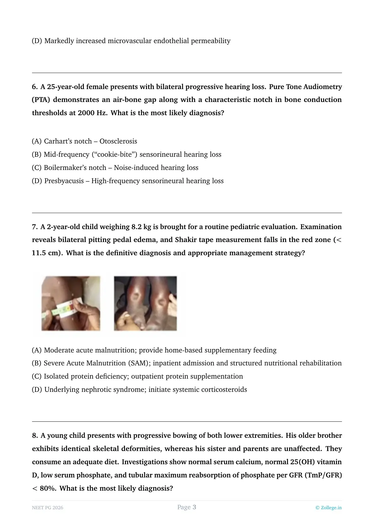
A 2-year-old child weighing 8.2 kg is brought for a routine pediatric evaluation. Examination reveals bilateral pitting pedal edema, and Shakir tape measurement falls in the red zone ($<$ 11.5 cm). What is the definitive diagnosis and appropriate management strategy?

View Solution
Step 1: Understanding the Question:
The child is 2 years old and presents with two major physical findings: bilateral pitting pedal edema and a Mid-Upper Arm Circumference (MUAC), measured by Shakir tape, of less than 11.5 cm (red zone). We must diagnose the severity of malnutrition and decide on the management setting.
Step 2: Key Formula or Approach:
According to WHO and National Health Mission (India) guidelines, Severe Acute Malnutrition (SAM) in children 6–59 months is diagnosed if: 1. MUAC $<$ 11.5 cm OR 2. Weight-for-Height/Length Z-score $<$ -3 SD OR 3. Presence of bilateral pitting edema of nutritional origin.
Step 3: Detailed Explanation:
1. Diagnosis: This child satisfies two criteria for SAM: the MUAC is in the red zone ($<$ 11.5 cm) and there is bilateral pitting edema. The presence of edema classifies this as "Edematous SAM" (clinically known as Kwashiorkor).
2. Management Strategy: While some children with "uncomplicated" SAM (good appetite, no medical issues) can be managed in the community (Outpatient Therapeutic Program), the presence of bilateral pitting edema is considered a major complication. Edematous children have high metabolic instability, risk of electrolyte imbalances (like hypokalemia), and a higher risk of sudden death. Therefore, they require "Inpatient Admission" to a Nutritional Rehabilitation Center (NRC).
3. Rehabilitation Phases: Treatment involves stabilization (Phase 1) with F-75 milk to restore metabolic function without overloading the system, followed by a transition to F-100 or RUTF (Ready-to-Use Therapeutic Food) for catch-up growth.
Step 4: Final Answer:
The child has SAM with complications (edema) and must be admitted to an inpatient facility for structured nutritional rehabilitation.
A young child presents with progressive bowing of both lower extremities. His older brother exhibits identical skeletal deformities, whereas his sister and parents are unaffected. They consume an adequate diet. Investigations show normal serum calcium, normal 25(OH) vitamin D, low serum phosphate, and tubular maximum reabsorption of phosphate per GFR (TmP/GFR) $<$ 80%. What is the most likely diagnosis?
View Solution
Step 1: Understanding the Question:
A child has rickets despite a good diet and normal 25-hydroxyvitamin D levels. The biochemical hallmarks are "low serum phosphate" and "low renal phosphate reabsorption" (TmP/GFR $<$ 80%). The family history shows only brothers affected, suggesting an X-linked dominant inheritance (common for this condition).
Step 2: Key Formula or Approach:
Rickets is broadly divided into Calcipenic and Phosphopenic types. If Calcium and Vitamin D are normal but Phosphate is low due to kidney "wasting," the condition is a phosphopenic rickets, most commonly X-linked Hypophosphatemic Rickets (XLHR).
Step 3: Detailed Explanation:
1. Pathophysiology: XLHR is caused by a mutation in the PHEX gene. This leads to an excess of FGF-23 (a phosphatonin). FGF-23 does two things: it inhibits the NPT2a/c transporters in the proximal tubule (causing phosphate wasting) and it inhibits the 1-alpha-hydroxylase enzyme, preventing the formation of active Vitamin D (1,25-OH2D).
2. Interpretation of Investigations: - Normal 25(OH)D: Rules out nutritional Vitamin D deficiency.
- Low Phosphate + Low TmP/GFR: Proves the kidney is actively "leaking" phosphate instead of reabsorbing it (normal reabsorption should be $>$ 85-90%).
- Clinical Presentation: Bowing of legs (Rickets) occurs because phosphate is essential for hydroxyapatite formation and bone mineralization.
3. Differential Analysis: - Fanconi Syndrome: Would also show loss of glucose, amino acids, and bicarbonate in urine (not mentioned). - Vitamin D Dependent Rickets: Would typically show low serum calcium and low/abnormal Vitamin D metabolites.
Step 4: Final Answer:
The combination of rickets, normal Vitamin D levels, and isolated renal phosphate wasting (low TmP/GFR) is characteristic of X-linked Hypophosphatemic Rickets.
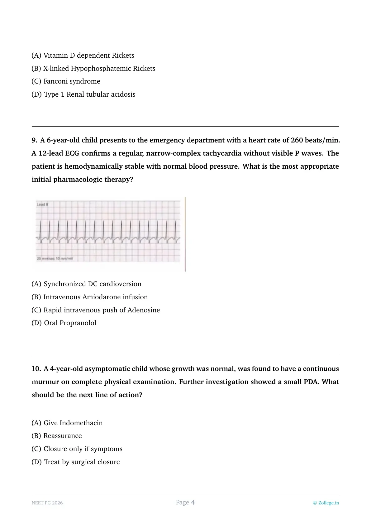
A 6-year-old child presents to the emergency department with a heart rate of 260 beats/min. A 12-lead ECG confirms a regular, narrow-complex tachycardia without visible P waves. The patient is hemodynamically stable with normal blood pressure. What is the most appropriate initial pharmacologic therapy?

View Solution
Step 1: Understanding the Question:
The child has a heart rate (260 bpm) that is way above the normal range for his age. The ECG shows "narrow-complex tachycardia" with "no visible P waves," which is the classic description of Supraventricular Tachycardia (SVT). The child is currently "hemodynamically stable" (normal blood pressure).
Step 2: Key Formula or Approach:
The PALS (Pediatric Advanced Life Support) management for SVT depends on stability:
1. Unstable (Shock/Hypotension): Immediate Synchronized Cardioversion.
2. Stable:
- Step 1: Vagal maneuvers (e.g., ice to face).
- Step 2: If vagal maneuvers fail, the first-line medication is Adenosine.
Step 3: Detailed Explanation:
1. Adenosine: It works by causing a transient, complete block at the Atrioventricular (AV) node. This "resets" the heart and breaks the re-entrant circuit that usually maintains SVT.
2. Administration: It has an extremely short half-life ($<$ 10 seconds). Therefore, it must be given as a "Rapid IV push" followed immediately by a rapid saline flush, ideally through a large-bore IV close to the heart.
3. Why not other options?
- Synchronized Cardioversion: Used only if the patient is unstable or if medications fail.
- Amiodarone: Used for resistant cases or ventricular arrhythmias; it is not first-line for narrow-complex SVT.
- Oral Propranolol: Acute arrhythmias require IV medication; oral drugs take too long to act.
Step 4: Final Answer:
For a hemodynamically stable child in SVT, the most appropriate initial pharmacologic therapy is a rapid IV push of Adenosine.
A 4-year-old asymptomatic child whose growth was normal, was found to have a continuous murmur on complete physical examination. Further investigation showed a small PDA. What should be the next line of action?
View Solution
Step 1: Understanding the Question:
The patient is a 4-year-old child with a "continuous murmur" and a confirmed small Patent Ductus Arteriosus (PDA). Although the child is currently asymptomatic, we must decide whether to monitor or intervene.
Step 2: Key Formula or Approach:
The natural history of PDA in children beyond infancy includes the risk of infective endarteritis and progressive left heart volume overload. If a PDA is "audible" (produces a murmur), clinical guidelines generally recommend closure regardless of the presence of symptoms.
Step 3: Detailed Explanation:
1. Rationale for Treatment: Even a "small" PDA is not benign. The turbulent flow across the ductus can cause endothelial damage, making it a prime site for Infective Endarteritis. Furthermore, even a small left-to-right shunt can lead to chamber enlargement and eventual heart failure or pulmonary hypertension in adulthood.
2. Method of Closure: While the question says "surgical closure," in modern practice, this is often done via Percutaneous Transcatheter Closure (using coils or occluders). However, the underlying principle is that "closure" (intervention) is the correct management.
3. Why not other options? - Indomethacin/Ibuprofen: These prostaglandin inhibitors only work to close a PDA in the first few days/weeks of life (neonatal period). They are ineffective in a 4-year-old. - Reassurance/Closure only if symptomatic: This is incorrect because waiting for symptoms means waiting for heart failure or endarteritis to occur, which is not standard of care for an audible PDA.
Step 4: Final Answer:
Because the PDA is clinically audible (continuous murmur), definitive treatment via closure is recommended to prevent long-term complications.
A 6-year-old child presented with hematuria and edema 2 weeks after an episode of sore throat. Light microscopy shows diffuse infiltration of the glomeruli by neutrophils. What is the most probable diagnosis?
View Solution
Step 1: Understanding the Question:
A 6-year-old child presents with signs of nephritic syndrome (hematuria and edema). There is a crucial history of a preceding sore throat (pharyngitis) with a "latency period" of 2 weeks. Histology shows an "exudative" pattern (neutrophil infiltration).
Step 2: Key Formula or Approach:
The combination of a post-infectious timeline (1–3 weeks for pharyngitis), clinical nephritic syndrome, and diffuse glomerular infiltration by neutrophils is the classic presentation of Acute Post-Streptococcal Glomerulonephritis (APSG/PSGN).
Step 3: Detailed Explanation:
1. Pathogenesis: PSGN is a Type III hypersensitivity reaction. Immune complexes (containing streptococcal antigens like SpeB) deposit in the glomeruli, activating the complement system and recruiting inflammatory cells.
2. Histology: On light microscopy, the glomeruli appear enlarged and "hypercellular." This hypercellularity is due to both the proliferation of resident cells and an "exudate" of inflammatory cells, primarily neutrophils and monocytes.
3. Differential Analysis: - RPGN: Characterized by "crescents" in Bowman's space, not just neutrophil infiltration. - Minimal Change Disease: Presents with nephrotic syndrome (massive proteinuria), not hematuria, and appears normal on light microscopy. - MPGN: Shows a "tram-track" appearance of the basement membrane and a more chronic course.
Step 4: Final Answer:
The clinical history of post-pharyngeal hematuria and the histological finding of neutrophilic infiltration point definitively to Acute Post Streptococcal Glomerulonephritis.
A 14-year-old boy developed seizures while on vacation. Serum calcium was 6.5 mg/dL and PTH was 218 pg/ml. What is the diagnosis?
View Solution
Step 1: Understanding the Question:
The patient has symptomatic hypocalcemia (seizures and Calcium = 6.5 mg/dL; normal is 8.5–10.5). Crucially, the Parathyroid Hormone (PTH) level is significantly elevated (218 pg/ml; normal is 10–65).
Step 2: Key Formula or Approach:
To differentiate between types of parathyroid disorders, look at the relationship between Calcium and PTH: - Low Ca + Low PTH = Primary Hypoparathyroidism. - Low Ca + High PTH = Either Secondary Hyperparathyroidism (e.g., Vitamin D deficiency) or Target Organ Resistance to PTH (Pseudohypoparathyroidism).
Step 3: Detailed Explanation:
1. Pseudohypoparathyroidism (PHP): This is a genetic condition (mutation in the GNAS1 gene) where the body's tissues (especially the kidneys) are resistant to the action of PTH. Even though the parathyroid glands are healthy and producing plenty of PTH, the "message" isn't received. This results in the biochemical paradox of hypocalcemia (due to lack of PTH effect) and secondary high PTH (as the glands try to compensate).
2. Differential Analysis:
- Primary Hypoparathyroidism: Would show low PTH because the gland itself is failing.
- Pseudopseudohypoparathyroidism (PPHP): This patient has the physical phenotype (short stature, short 4th metacarpal) but has normal Calcium and PTH levels.
- Hyperparathyroidism: Usually presents with *high* Calcium.
Step 4: Final Answer:
The combination of low serum calcium and elevated PTH indicates end-organ resistance, which is diagnostic of Pseudohypoparathyroidism.
A patient underwent surgery in the posterior triangle of the neck 3 weeks ago. He now presents with difficulty elevating the arm above shoulder level and difficulty shrugging the shoulder. Which of the following nerve is most likely injured?
View Solution
Step 1: Understanding the Question:
The patient had surgery in the posterior triangle of the neck. Post-operatively, he cannot shrug his shoulder and has trouble with high arm elevation. These symptoms correspond to a weakness of the Trapezius muscle.
Step 2: Key Formula or Approach:
The Spinal Accessory Nerve (CN XI) has a very superficial course across the posterior triangle of the neck (resting on the levator scapulae). It is the most commonly injured nerve during minor surgeries in this area (like lymph node biopsies).
Step 3: Detailed Explanation:
1. Trapezius Function: The trapezius muscle is responsible for shrugging the shoulders (upper fibers) and helps in the upward rotation of the scapula, which is essential for abducting the arm above 90 degrees.
2. Clinical Sign: Injury to CN XI leads to drooping of the shoulder, winging of the scapula (lateral), and the specific inability to shrug or perform overhead abduction.
3. Differential Analysis:
- Long thoracic nerve: Supplies the Serratus Anterior. Injury causes "medial" winging and difficulty with forward punching, but shrugging is usually preserved.
- Suprascapular nerve: Supplies Supraspinatus and Infraspinatus; injury affects the initiation of abduction and external rotation, not shrugging.
Step 4: Final Answer:
Given the surgical location (posterior triangle) and the clinical deficit (shrugging), the Spinal Accessory nerve is the site of injury.
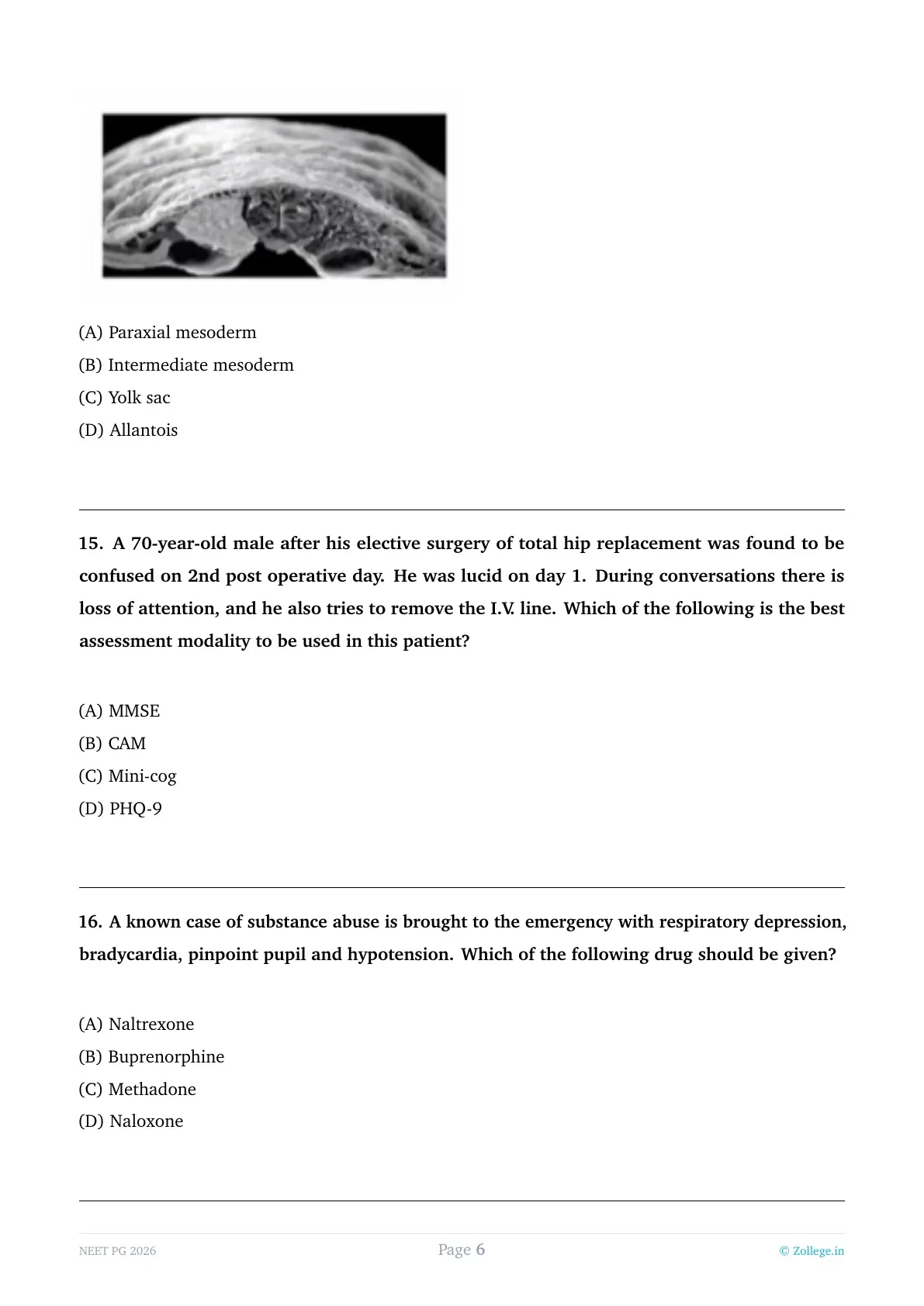
Kidney develops from which of the following parts?

View Solution
Step 1: Understanding the Question:
The question asks for the embryological origin of the kidney (part of the urogenital system).
Step 2: Key Formula or Approach:
The intraembryonic mesoderm is divided into three parts: Paraxial (somites), Intermediate, and Lateral plate mesoderm. The entire urinary system and most of the genital system develop from the intermediate mesoderm.
Step 3: Detailed Explanation:
1. Intermediate Mesoderm: During folding of the embryo, this mesoderm forms a longitudinal ridge called the urogenital ridge. It gives rise to the three successive kidney systems:
- Pronephros: Vestigial and disappears.
- Mesonephros: Functions briefly in the first trimester.
- Metanephros: Forms the permanent kidney (the excretory part).
2. Metanephros Components: The permanent kidney actually has a dual origin within the intermediate mesoderm:
- Ureteric bud: Gives rise to the collecting system (ureter, pelvis, calyces, collecting tubules).
- Metanephric blastema: Gives rise to the nephrons (Bowman's capsule to DCT).
3. Other Options: Paraxial mesoderm forms bone and muscle of the trunk. Yolk sac is involved in early hematopoiesis and germ cell origin. Allantois contributes to the formation of the urachus and bladder.
Step 4: Final Answer:
The kidney develops from the intermediate mesoderm.
A 70-year-old male after his elective surgery of total hip replacement was found to be confused on 2nd post operative day. He was lucid on day 1. During conversations there is loss of attention, and he also tries to remove the I.V. line. Which of the following is the best assessment modality to be used in this patient?
View Solution
Step 1: Understanding the Question:
An elderly post-operative patient shows acute onset of confusion, fluctuating course (lucid on day 1, confused on day 2), and "loss of attention" (inattention). He is also agitated (pulling IV lines). This is a classic presentation of Delirium.
Step 2: Key Formula or Approach:
When diagnosing delirium, the "Gold Standard" bedside screening and assessment tool is the Confusion Assessment Method (CAM).
Step 3: Detailed Explanation:
1. CAM (Confusion Assessment Method): It is specifically designed to identify delirium by looking for four features: (1) Acute onset and fluctuating course, (2) Inattention, (3) Disorganized thinking, and (4) Altered level of consciousness. A diagnosis of delirium requires features 1 and 2, plus either 3 or 4.
2. Why not other options? - MMSE (Mini-Mental State Exam): Used primarily to screen for Dementia (chronic cognitive loss). It is poor at distinguishing delirium from dementia and does not assess the fluctuating nature or inattention well.
- Mini-cog: A very quick screening tool for dementia (3-item recall and clock drawing).
- PHQ-9: This is a screening tool for Depression, not cognitive impairment or delirium.
Step 4: Final Answer:
The best modality for assessing a patient with suspected post-operative delirium is the CAM.
A known case of substance abuse is brought to the emergency with respiratory depression, bradycardia, pinpoint pupil and hypotension. Which of the following drug should be given?
View Solution
Step 1: Understanding the Question:
The patient presents with the classic clinical triad of opioid overdose: respiratory depression, altered mental status, and miosis (pinpoint pupils). Associated signs like bradycardia and hypotension further support severe opioid toxicity. We need to identify the immediate life-saving pharmacological intervention to reverse these effects.
Step 2: Key Formula or Approach:
In the emergency management of acute opioid poisoning, the priority is maintaining the airway and reversing the respiratory depression. This is achieved using a pure competitive opioid antagonist that has a rapid onset of action when administered intravenously.
Step 3: Detailed Explanation:
1. Naloxone: This is a pure opioid antagonist that has a high affinity for the $mu$-opioid receptors. When administered intravenously, it acts within 1–2 minutes to displace opioid molecules from their receptors, thereby reversing all physiological effects of the opioid, including life-threatening respiratory depression and sedation. It is the gold standard for acute overdose management in the emergency department.
2. Naltrexone: While also an antagonist, it has a much longer half-life and is primarily used for the long-term maintenance of opioid-free states (relapse prevention) and the treatment of alcohol dependence. It is generally administered orally and has a slow onset of action, making it inappropriate for an acute overdose emergency.
3. Methadone and Buprenorphine: These are opioid agonists (Methadone is a full agonist; Buprenorphine is a partial agonist). Administering these to a patient already suffering from an overdose would exacerbate the respiratory depression and could lead to a fatal outcome. These drugs are used for detoxification and long-term maintenance therapy in controlled clinical settings.
Step 4: Final Answer:
Naloxone is the definitive drug of choice for the immediate reversal of life-threatening respiratory depression and CNS depression in an opioid overdose.
A 24-year-old male patient with bipolar disorder was taking lithium 900 mg/ day and quetiapine 50mg/day. He started having tremors, dysarthria and ataxia. Which of the following is correct statement related to the patient?
View Solution
Step 1: Understanding the Question:
A patient diagnosed with bipolar disorder and maintained on Lithium and a low dose of Quetiapine develops new neurological symptoms: tremors, dysarthria (slurred speech), and ataxia (loss of coordination). We must determine the most likely cause of these symptoms in the context of his medication regimen.
Step 2: Key Formula or Approach:
Lithium has a very narrow therapeutic index (therapeutic range is 0.6–1.2 mEq/L). While mild side effects occur at therapeutic levels, moderate-to-severe neurological symptoms are characteristic of Lithium toxicity, which can occur even if the prescribed dose hasn't changed (due to factors like dehydration or drug interactions).
Step 3: Detailed Explanation:
1. Lithium Toxicity: As serum lithium levels rise above 1.5 mEq/L, toxicity manifests primarily through the Central Nervous System. Early signs include "coarse" tremors (as opposed to the "fine" tremors seen at therapeutic levels). As levels reach the 2.0–2.5 mEq/L range, patients develop ataxia, dysarthria, muscle fasciculations, and severe drowsiness. The presence of ataxia and slurred speech in this patient is a classic clinical "red flag" for toxicity.
2. Extrapyramidal Symptoms (EPS): Quetiapine is a second-generation antipsychotic with a very low affinity for $D_2$ receptors, making EPS very rare, especially at the low dose of 50 mg (usually used for sleep or mood stabilization). EPS typically presents as dystonia or parkinsonism, not ataxia.
3. Essential Tremors: These are typically postural/action tremors that are not associated with dysarthria or ataxia.
Step 4: Final Answer:
The constellation of coarse tremors, dysarthria, and ataxia in a patient taking Lithium is diagnostic of Lithium-induced toxicity until proven otherwise by a serum Lithium assay.
A 25-year-old male is presents with long standing pattern of social withdrawal. He has odd and eccentric behavior and hold some peculiar beliefs. Which of the following personality traits are seen here?
View Solution
Step 1: Understanding the Question:
The patient displays a chronic pattern of social isolation, but significantly, he also exhibits "odd and eccentric behavior" and "peculiar beliefs." We need to distinguish between the various personality disorders that involve social withdrawal.
Step 2: Key Formula or Approach:
Psychiatry categorizes personality disorders into three clusters. Cluster A (the "odd/eccentric" cluster) includes Paranoid, Schizoid, and Schizotypal.
- Schizoid: Socially isolated + Content with being alone.
- Schizotypal: Socially isolated + "Odd" thoughts/appearance/magical thinking.
Step 3: Detailed Explanation:
1. Schizotypal Personality Disorder: These individuals are characterized by acute discomfort in social relationships and a reduced capacity for close ones. However, the hallmark is cognitive or perceptual distortions. This includes "magical thinking" (peculiar beliefs in telepathy, clairvoyance, or "sixth sense"), unusual speech patterns, and eccentric behavior or appearance. The description in the question fits these criteria perfectly.
2. Schizoid Personality Disorder: These patients are "loners" by choice and show emotional coldness or detachment. They do not have the odd beliefs, magical thinking, or eccentricities that define the schizotypal personality.
3. Anxious/Avoidant: These individuals want social contact but are paralyzed by the fear of being criticized or rejected. They do not exhibit odd or peculiar behaviors.
4. Antisocial: This is a Cluster B disorder characterized by a lack of empathy and a disregard for social norms and the rights of others, which is not what is described here.
Step 4: Final Answer:
The combination of social withdrawal with "odd/eccentric" behavior and "peculiar beliefs" is the classic definition of Schizotypal personality.
An adult male patient suffered head injury and during recovery, the patient was drinking 8liters of water per day, with intense third and make large dilute urine. There is no diabetes mellitus. Which hypothalamic nucleus and descending fibers are damaged?
View Solution
Step 1: Understanding the Question:
Following a head injury, the patient develops extreme polyuria (8L of water) and polydipsia (intense thirst) with dilute urine (low specific gravity). In the absence of diabetes mellitus, this clinical picture is diagnostic of Diabetes Insipidus (DI). In a post-trauma setting, this is almost always Central DI.
Step 2: Key Formula or Approach:
Central Diabetes Insipidus is caused by a deficiency of Antidiuretic Hormone (ADH), also known as Arginine Vasopressin. To solve this, we must identify which part of the brain synthesizes ADH and the pathway through which it reaches the circulation.
Step 3: Detailed Explanation:
1. Synthesis of ADH: ADH is a peptide hormone synthesized by the magnocellular neurons located in the Supraoptic nucleus and the Paraventricular nucleus of the hypothalamus. While the supraoptic nucleus is the primary producer of ADH (and paraventricular produces more Oxytocin), both nuclei contribute to ADH production.
2. Transport and Storage: Once synthesized, the hormone is transported down the axons of these neurons, which form the hypothalamo-hypophyseal tract (the "descending fibers"). These fibers pass through the pituitary stalk to the posterior pituitary (neurohypophysis), where the hormone is stored and eventually released into the blood.
3. Pathophysiology of Injury: A head injury or skull base fracture can shear these axons or damage the nuclei themselves. This stops the production and/or release of ADH. Without ADH, the collecting ducts of the kidney become impermeable to water, leading to the excretion of massive volumes of dilute urine.
Step 4: Final Answer:
The destruction of the Paraventricular and Supraoptic nuclei or their descending tracts in the pituitary stalk leads to the development of Central Diabetes Insipidus.
An adult women is investigated for mild weakness, fatigue and occasional carpopedal spasm. Laboratory studies reveals, hypokalemia, metabolic alkalosis and high calcium in the urine. In which part of the nephron the defective channel is present?
View Solution
Step 1: Understanding the Question:
The patient has a classic biochemical profile: hypokalemia and metabolic alkalosis. Clinically, she has "carpopedal spasms," which usually suggest hypocalcemia (or a decrease in ionized calcium due to alkalosis). The laboratory hallmark that differentiates this case is high calcium in the urine (hypercalciuria).
Step 2: Key Formula or Approach:
The two primary hereditary tubulopathies that cause hypokalemic metabolic alkalosis with normal-to-low blood pressure are Bartter Syndrome and Gitelman Syndrome.
- Bartter Syndrome: Mimics Loop Diuretics; associated with Hypercalciuria.
- Gitelman Syndrome: Mimics Thiazide Diuretics; associated with Hypocalciuria.
Step 3: Detailed Explanation:
1. Localization to the TAL: Bartter Syndrome is caused by mutations in the transporters of the Thick Ascending Limb (TAL) of the Loop of Henle, such as the NKCC2 (Na-K-2Cl cotransporter), ROMK (potassium channel), or CLC-Kb (chloride channel).
2. Mechanism of Hypercalciuria: Under normal conditions, the reabsorption of K+ through ROMK creates a positive luminal potential in the TAL. This voltage is the driving force for the "paracellular" (between cells) reabsorption of divalent cations like Calcium and Magnesium. When the TAL transporters are defective (as in Bartter syndrome or with Furosemide use), this positive potential is not generated, Calcium is not reabsorbed, and it is excreted in the urine.
3. Why Alkalosis? The failure of the TAL to reabsorb sodium leads to increased sodium delivery to the distal nephron. The body attempts to reclaim this sodium in exchange for Potassium and Hydrogen ions, leading to hypokalemia and metabolic alkalosis.
Step 4: Final Answer:
The presence of hypercalciuria (high urine calcium) localizes the defect to the Thick Ascending Limb of the Loop of Henle.
A 28-year-old man presents with a painless testicular mass. On gross examination, the tumor is well-circumscribed, homogeneous, and gray-white, with a soft, fleshy appearance and minimal hemorrhage or necrosis. Histological examination shows sheets and nests of uniform polygonal cells with abundant clear cytoplasm containing glycogen and prominent nucleoli, separated by delicate fibrous septa infiltrated by lymphocytes. Immunohistochemistry shows tumor cells that are positive for CD117 (c-KIT) and OCT3/4. What is the most likely diagnosis?
View Solution
Step 1: Understanding the Question:
The patient is a young adult male with a painless testicular mass. The description provided includes gross findings (homogeneous, fleshy, no necrosis), microscopic findings (clear cells, fibrous septa with lymphocytes), and specific immunohistochemistry (IHC) markers (CD117, OCT3/4). We need to correlate these findings with a specific germ cell tumor.
Step 2: Key Formula or Approach:
In testicular pathology, the "classic" description of a seminoma is a "fleshy, gray-white mass" that histologically shows "clear cells separated by lymphocytic septa." IHC markers are definitive: Seminomas are characteristically positive for CD117 (c-KIT), OCT3/4, and SALL4.
Step 3: Detailed Explanation:
1. Seminoma: This is the most common germ cell tumor in men, typically occurring in the 3rd and 4th decades. Grossly, it is remarkably "pure" and homogeneous, lacking the hemorrhage and necrosis seen in non-seminomatous germ cell tumors. Microscopic examination shows large, uniform cells with clear cytoplasm (due to high glycogen content) and distinct cell borders. The presence of lymphocytes within the fibrous septa is a hallmark diagnostic feature.
2. Immunohistochemistry: CD117 (c-KIT) is a membrane-bound receptor tyrosine kinase that is highly sensitive for seminoma. OCT3/4 is a marker of pluripotency also found in seminomas and embryonal carcinomas. However, the combination of CD117(+) and the lymphocytic infiltrate is specific to seminoma among the options.
3. Differential Analysis: - Embryonal carcinoma: Grossly shows extensive hemorrhage and necrosis. Cells are pleomorphic, forming glands or sheets, and are typically CD117 negative. - Yolk sac tumor: Characterized by Schiller-Duval bodies and positivity for Alpha-Fetoprotein (AFP). - Choriocarcinoma: Shows syncytiotrophoblasts and cytotrophoblasts with high levels of Beta-hCG and significant hemorrhage.
Step 4: Final Answer:
The homogeneous fleshy appearance, lymphocytic infiltrate in septa, and CD117 positivity are diagnostic of a Seminoma.
A 65-year-old chronic smoker presents with cough, weight loss, and progressive dyspnea. Chest imaging reveals a centrally located hilar lung mass. Histological examination shows sheets of small round blue cells with scant cytoplasm, hyperchromatic nuclei, a high nuclear-to-cytoplasmic ratio, nuclear molding, frequent mitotic figures, and areas of necrosis. Immunohistochemistry shows tumor cells that are positive for synaptophysin. What is the most likely diagnosis?
View Solution
Step 1: Understanding the Question:
The patient is an elderly smoker with a central (hilar) lung mass. The histological description is very specific: "small round blue cells," "nuclear molding," and "high N:C ratio." Additionally, the tumor is positive for "synaptophysin," which is a neuroendocrine marker.
Step 2: Key Formula or Approach:
Lung cancers are divided into Small Cell Lung Cancer (SCLC) and Non-Small Cell Lung Cancer (NSCLC). Central masses in smokers are typically either Small Cell or Squamous Cell. The histological finding of "small blue cells" and "neuroendocrine markers" (synaptophysin, chromogranin, CD56) is pathognomonic for Small Cell Carcinoma.
Step 3: Detailed Explanation:
1. Small Cell Carcinoma (SCLC): This is a highly aggressive tumor strongly linked to smoking. It typically arises centrally near the hila. The cells are derived from neuroendocrine cells (Kulchitsky cells) of the bronchial epithelium.
2. Microscopic Features: The cells are small (about 2-3 times the size of a lymphocyte), have very little cytoplasm (scant), and hyperchromatic nuclei. Nuclear molding occurs because the cells are so tightly packed that their nuclei press against and "mold" to each other. Frequent mitoses and "Azzopardi effect" (DNA smearing from necrotic cells in vessel walls) are also common.
3. Immunohistochemistry: Because it is a neuroendocrine tumor, it expresses markers like Synaptophysin, Chromogranin A, and CD56.
4. Differential Analysis:
- Squamous cell carcinoma: Also central and seen in smokers, but characterized by keratin pearls and intercellular bridges.
- Adenocarcinoma: Usually peripheral and shows gland formation or mucin production; it is the most common lung cancer in non-smokers.
Step 4: Final Answer:
The combination of a hilar mass in a smoker, small round blue cells with nuclear molding, and synaptophysin positivity identifies the tumor as Small Cell Carcinoma.
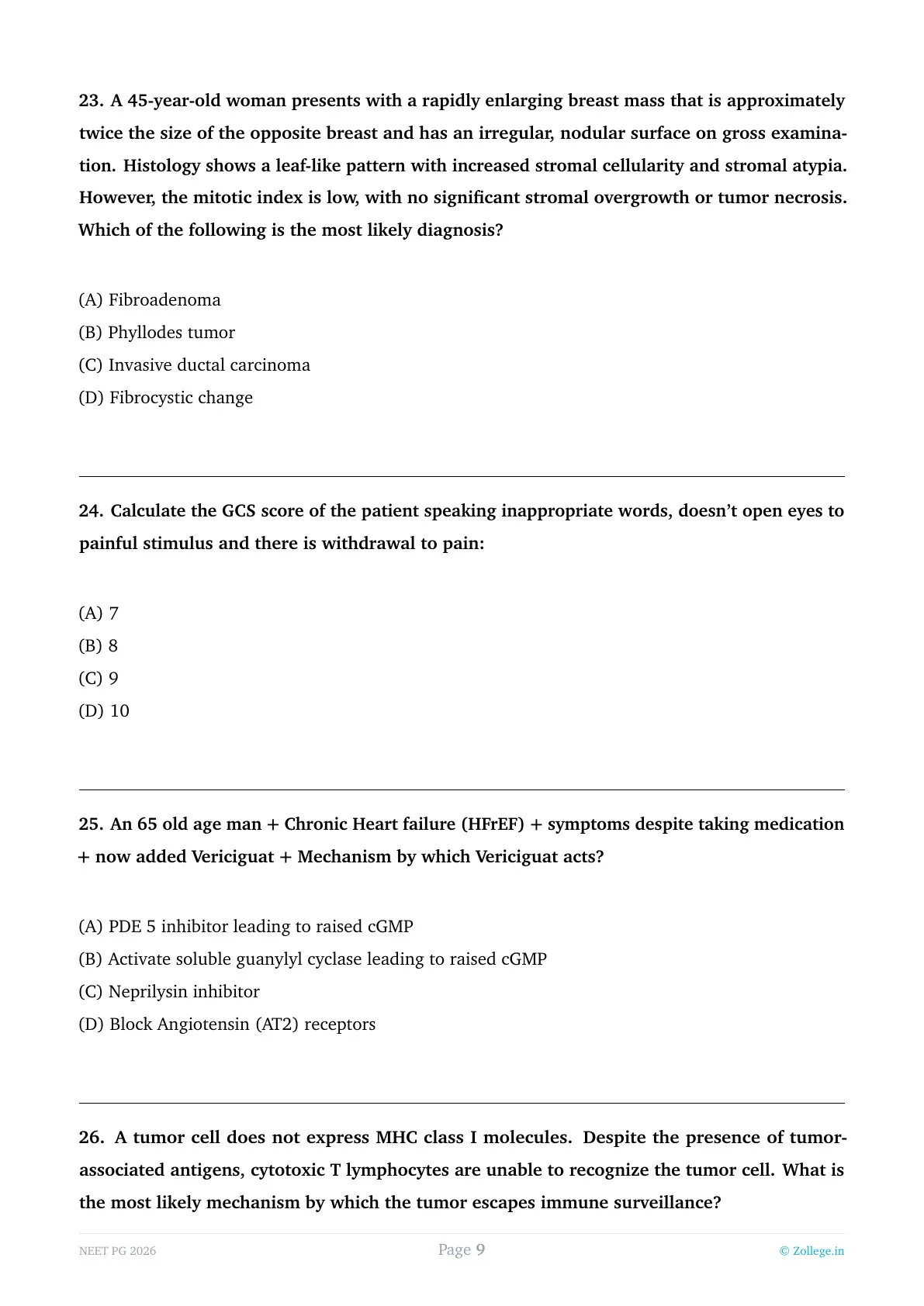
A 45-year-old woman presents with a rapidly enlarging breast mass that is approximately twice the size of the opposite breast and has an irregular, nodular surface on gross examination. Histology shows a leaf-like pattern with increased stromal cellularity and stromal atypia. However, the mitotic index is low, with no significant stromal overgrowth or tumor necrosis. Which of the following is the most likely diagnosis?
View Solution
Step 1: Understanding the Question:
The patient is a middle-aged woman with a very large, rapidly growing breast mass. The histological hallmark mentioned is a "leaf-like pattern" and "increased stromal cellularity." We need to distinguish this from other fibroepithelial lesions like fibroadenoma.
Step 2: Key Formula or Approach:
Breast lesions with both epithelial and stromal components are "fibroepithelial lesions." When the stroma becomes hypercellular and grows into "leaf-like" (phyllodes) projections, the diagnosis is a Phyllodes tumor.
Step 3: Detailed Explanation:
1. Phyllodes Tumor: These tumors originate from the intralobular stroma. They are distinguished from fibroadenomas by their much higher stromal cellularity and the characteristic "leaf-like" architectural pattern (cleft-like spaces lined by epithelium).
2. Grading: They are classified as Benign, Borderline, or Malignant based on stromal atypia, mitotic activity, and stromal overgrowth. The case described has "increased stromal cellularity and atypia" but a "low mitotic index" and "no stromal overgrowth," suggesting a Benign or Low-grade Phyllodes tumor.
3. Clinical Behavior: Unlike fibroadenomas, Phyllodes tumors tend to grow rapidly and reach large sizes (often $>5$ cm). They have a tendency for local recurrence if not excised with wide margins.
4. Differential Analysis:
- Fibroadenoma: Usually smaller, with less cellular stroma and no "leaf-like" architecture.
- Invasive Ductal Carcinoma: Characterized by malignant epithelial cells invading the stroma, not a proliferation of the stroma itself.
Step 4: Final Answer:
The "leaf-like pattern" and increased stromal cellularity in a large, rapidly growing breast mass are pathognomonic for a Phyllodes tumor.
Calculate the GCS score of the patient speaking inappropriate words, doesn’t open eyes to painful stimulus and there is withdrawal to pain:
View Solution
Step 1: Understanding the Question:
We are required to calculate the total Glasgow Coma Scale (GCS) score based on three clinical parameters: eye opening, verbal response, and motor response.
Step 2: Key Formula or Approach:
The GCS formula is: Total Score = E + V + M
1. Eye Opening (E): 4 (Spontaneous), 3 (To sound), 2 (To pressure/pain), 1 (None).
2. Verbal Response (V): 5 (Oriented), 4 (Confused), 3 (Inappropriate words), 2 (Incomprehensible sounds), 1 (None).
3. Motor Response (M): 6 (Obeys commands), 5 (Localizing pain), 4 (Normal withdrawal), 3 (Abnormal flexion/decorticate), 2 (Extension/decerebrate), 1 (None).
Step 3: Detailed Explanation:
1. Eye Response (E): The patient "doesn't open eyes to painful stimulus." This indicates the lowest possible score for eyes. E = 1.
2. Verbal Response (V): The patient is "speaking inappropriate words." These are recognizable words but used in a random or exclamatory way, not forming meaningful sentences. V = 3.
3. Motor Response (M): There is "withdrawal to pain." This means the patient pulls the limb away from a painful stimulus but does not reach across the midline to stop the stimulus (which would be localizing). M = 4.
4. Calculation: Total GCS = $1 (E) + 3 (V) + 4 (M) = 8$.
Step 4: Final Answer:
The calculated GCS score for this patient is 8.
An 65 old age man + Chronic Heart failure (HFrEF) + symptoms despite taking medication + now added Vericiguat + Mechanism by which Vericiguat acts?
View Solution
Step 1: Understanding the Question:
A patient with Heart Failure with reduced Ejection Fraction (HFrEF) is still symptomatic despite standard therapy. A new drug, Vericiguat, is added. We need to identify its mechanism of action.
Step 2: Key Formula or Approach:
In heart failure, the Nitric Oxide (NO) - soluble Guanylyl Cyclase (sGC) - cyclic Guanosine Monophosphate (cGMP) pathway is impaired. cGMP is vital for myocardial and vascular relaxation. Vericiguat targets this pathway.
Step 3: Detailed Explanation:
1. Vericiguat: It is a first-in-class oral soluble guanylate cyclase (sGC) stimulator.
2. Mechanism: It acts by directly binding to and stimulating sGC, independently of nitric oxide (NO). It also sensitizes sGC to endogenous NO by stabilizing NO binding.
3. Downstream Effects: Activation of sGC leads to the conversion of GTP to cGMP. Increased levels of intracellular cGMP lead to smooth muscle relaxation (vasodilation), improved endothelial function, and reduced myocardial stiffness/fibrosis.
4. Clinical Use: It was approved (based on the VICTORIA trial) for use in patients with HFrEF following a recent worsening heart failure event (hospitalization or IV diuretics) to reduce the risk of cardiovascular death and heart failure hospitalization.
5. Differential Analysis: - PDE 5 inhibitors (like Sildenafil) prevent the breakdown of cGMP but do not stimulate its production directly like Vericiguat. - Neprilysin inhibitors (like Sacubitril in ARNI) prevent the degradation of natriuretic peptides.
Step 4: Final Answer:
Vericiguat acts by directly activating soluble guanylyl cyclase, which increases the production of cGMP.
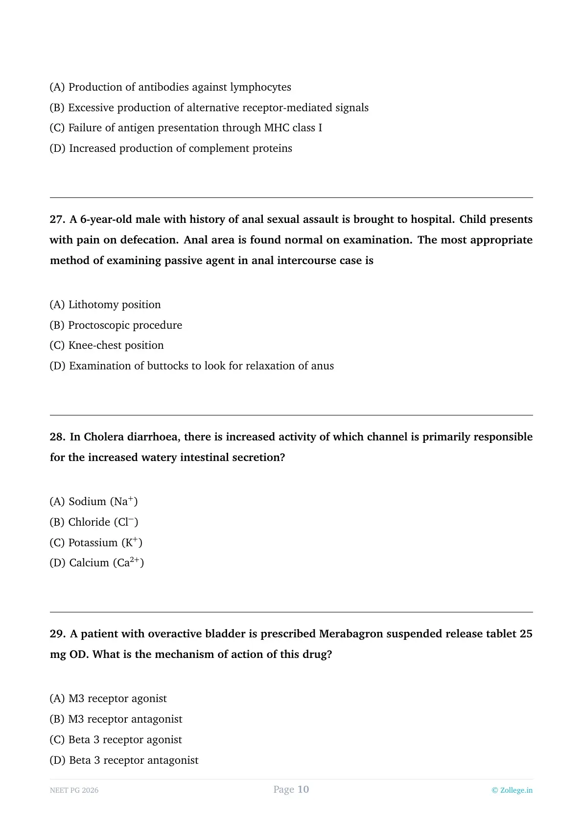
A tumor cell does not express MHC class I molecules. Despite the presence of tumor-associated antigens, cytotoxic T lymphocytes are unable to recognize the tumor cell. What is the most likely mechanism by which the tumor escapes immune surveillance?
View Solution
Step 1: Understanding the Question:
The question describes a scenario where a tumor cell has antigens (tumor-associated antigens) that should theoretically be recognized by the immune system, specifically by cytotoxic T lymphocytes (CD8+ T cells). However, the tumor cell has lost the expression of MHC class I molecules. We need to identify how this leads to immune escape.
Step 2: Key Formula or Approach:
The fundamental "rule" of T cell activation is MHC restriction. CD8+ Cytotoxic T Lymphocytes (CTLs) can only recognize antigens when they are processed into small peptides and presented on the cell surface within the groove of an MHC class I molecule.
Step 3: Detailed Explanation:
1. Role of MHC I: Every nucleated cell in the body expresses MHC class I. It serves as a "window" into the cell's internal protein environment. If a cell becomes cancerous, it produces abnormal proteins (antigens). These are chopped up and displayed on MHC I.
2. The Escape Mechanism: One of the most common ways tumors evade the immune system is by downregulating or completely losing the expression of MHC class I (often through mutations in the $beta_2$-microglobulin gene or TAP transporters).
3. Consequence: Without the MHC I "platform," the T-cell receptor (TCR) on the CD8+ lymphocyte has nothing to bind to. Even if the cell is full of "foreign" tumor antigens, the CTL remains "blind" to the cell's malignant state. This is a classic example of "Immune Editing" or "Immune Evasion."
4. Note on NK Cells: While the loss of MHC I protects the tumor from T cells, it theoretically makes them targets for Natural Killer (NK) cells, which follow the "missing self" hypothesis. However, tumors often develop additional mechanisms to inhibit NK cells as well.
Step 4: Final Answer:
The lack of MHC class I molecules results in the failure of antigen presentation, preventing cytotoxic T lymphocytes from recognizing and killing the tumor cell.
MHC 1 × CD8 = 8.
MHC 2 × CD4 = 8.
If you remove the MHC, the corresponding T-cell becomes functionally useless against that cell.
A 6-year-old male with history of anal sexual assault is brought to hospital. Child presents with pain on defecation. Anal area is found normal on examination. The most appropriate method of examining passive agent in anal intercourse case is
View Solution
Step 1: Understanding the Question:
In a case of suspected anal sexual assault (sodomy) where the victim is the "passive agent," a forensic medical examination is required. Even if the initial visual inspection seems "normal," specific maneuvers must be performed to check for signs of penetration or sphincter trauma.
Step 2: Key Formula or Approach:
Forensic examination of the anus involves looking for both "primary" signs (injuries, fissures, semen) and "secondary" signs (changes in sphincter tone). One of the most significant findings in cases of penetration is the loss of the normal anal reflex and tone.
Step 3: Detailed Explanation:
1. The Procedure: The victim is usually placed in the Knee-chest or lateral position. The examiner then gently but firmly separates the buttocks with both hands.
2. Reflex Anal Dilatation (RAD): In a normal individual, this action might cause the sphincter to contract. However, in a victim of anal penetration (especially if chronic or if there is significant recent trauma/stretching), the anus may relax or dilate instead of contracting. This is sometimes referred to as the "Reflex Anal Dilatation" or looking for the "funnel-shaped" appearance.
3. Significance: This relaxation or "patulousness" of the anus is a critical forensic finding for proving the act of penetration in a passive agent.
4. Why not others? While the Knee-chest (C) is a valid position, option (D) describes the actual method/maneuver used to elicit the forensic sign of sodomy. Proctoscopy (B) is generally used to look for internal injuries but is not the primary screening method for sphincter relaxation.
Step 4: Final Answer:
Separating the buttocks to observe the relaxation or reflex dilatation of the anal sphincter is the standard method to evaluate a suspected passive agent in sodomy cases.
In Cholera diarrhoea, there is increased activity of which channel is primarily responsible for the increased watery intestinal secretion?
View Solution
Step 1: Understanding the Question:
Cholera is characterized by "rice-water stools" (massive secretory diarrhea). We need to identify the specific ion channel in the intestinal epithelial cells that is overactivated to drive this fluid loss.
Step 2: Key Formula or Approach:
The pathophysiology of Vibrio cholerae involves the Cholera Toxin (CT), which increases intracellular cAMP. Increased cAMP acts as a second messenger that opens specific apical membrane channels.
Step 3: Detailed Explanation:
1. Mechanism of Toxin: The A-subunit of the cholera toxin enters the enterocyte and causes permanent ADP-ribosylation of the Gs protein. This keeps Adenylate Cyclase in the "ON" position.
2. cAMP Elevation: This leads to a massive rise in intracellular cAMP.
3. Channel Activation: cAMP directly activates the CFTR (Cystic Fibrosis Transmembrane conductance Regulator) protein, which is a Chloride channel located on the apical (luminal) surface of the crypt cells.
4. Fluid Shift: As Chloride ions are pumped into the intestinal lumen, an electrochemical gradient is created. Sodium (Na$^+$) and water follow paracellularly (between cells) into the lumen to maintain neutrality and osmotic balance. This results in the massive secretion of isotonic fluid, which exceeds the reabsorptive capacity of the colon, leading to diarrhea.
Step 4: Final Answer:
The primary channel responsible for the watery secretion in Cholera is the Chloride (Cl$^-$) channel (CFTR).
A patient with overactive bladder is prescribed Merabagron suspended release tablet 25 mg OD. What is the mechanism of action of this drug?
View Solution
Step 1: Understanding the Question:
The drug mentioned is Mirabegron (spelled Merabagron in the question). It is used to treat "Overactive Bladder" (OAB), a condition characterized by urgency, frequency, and nocturia. We need to identify its pharmacological class.
Step 2: Key Formula or Approach:
Traditionally, OAB was treated with anticholinergics (M3 antagonists like Oxybutynin). Mirabegron represents a newer class of drugs that work by stimulating the sympathetic (relaxation) pathway of the bladder rather than blocking the parasympathetic (contraction) pathway.
Step 3: Detailed Explanation:
1. Mechanism: Mirabegron is a potent and selective Beta-3 (\(\beta_3\)) adrenergic receptor agonist.
2. Action on the Bladder: These \(\beta_3\) receptors are primarily located on the detrusor muscle. When Mirabegron binds to these receptors, it induces detrusor muscle relaxation during the "storage phase" of the micturition cycle.
3. Clinical Effect: By relaxing the bladder during filling, it increases bladder capacity and reduces the frequency of involuntary detrusor contractions, thereby alleviating the symptoms of urgency and frequency.
4. Advantages: Unlike M3 antagonists, \(\beta_3\) agonists do not cause the "anti-cholinergic" side effects like dry mouth, blurred vision, or constipation, making them better tolerated in many patients.
Step 4: Final Answer:
Mirabegron is a Beta-3 receptor agonist.
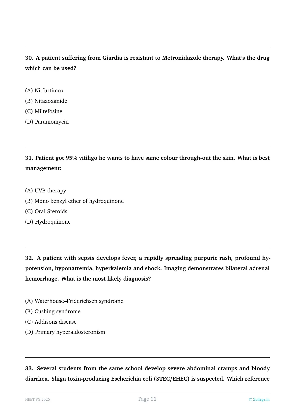
A patient suffering from Giardia is resistant to Metronidazole therapy. What’s the drug which can be used?
View Solution
Step 1: Understanding the Question:
The patient has Giardiasis (caused by the protozoan Giardia lamblia) but has not responded to the standard first-line treatment, Metronidazole. We need to identify an effective alternative.
Step 2: Key Formula or Approach:
Metronidazole and Tinidazole are nitroimidazoles. When resistance occurs, drugs from other classes or newer broad-spectrum antiparasitics are considered.
Step 3: Detailed Explanation:
1. Nitazoxanide: This is a broad-spectrum thiazolide anti-infective agent. It is highly effective against Giardia and is often the preferred choice for metronidazole-resistant cases or in pediatric patients (available as a liquid). It works by inhibiting the pyruvate:ferredoxin oxidoreductase (PFOR) enzyme, which is essential for anaerobic energy metabolism in the parasite.
2. Other Alternatives: Other drugs used for Giardia include Tinidazole (more potent than metronidazole), Albendazole, or Quinacrine.
3. Evaluation of Options:
- Nifurtimox: Used for Chagas disease (T. cruzi).
- Miltefosine: Primarily used for Leishmaniasis.
- Paromomycin: An aminoglycoside that is poorly absorbed from the gut. While it can be used for Giardia, it is generally reserved for pregnant women in the first trimester because it lacks systemic absorption and fetal risk. It is less effective than Nitazoxanide for resistant cases in general.
Step 4: Final Answer:
Nitazoxanide is an effective and common alternative for Metronidazole-resistant Giardiasis.
1st line: Metronidazole / Tinidazole.
Alternatives: Nitazoxanide / Albendazole.
In Pregnancy: Paromomycin (especially in the 1st trimester).
Patient got 95% vitiligo he wants to have same colour through-out the skin. What is best management:
View Solution
Step 1: Understanding the Question:
The patient has near-total vitiligo (95% of body surface area). At this stage, repigmentation of the affected 95% is extremely difficult and unlikely to be successful or aesthetically uniform. The patient's goal is to have a "uniform" skin color.
Step 2: Key Formula or Approach:
In "Vitiligo Universalis" (involving $>$ 50-80% of the body), the standard management strategy is not repigmentation, but depigmentation of the remaining small islands of normally pigmented skin to achieve a uniform, white appearance.
Step 3: Detailed Explanation:
1. Depigmentation Therapy: The gold standard agent for permanent depigmentation is Monobenzyl ether of hydroquinone (MBEH), usually in a 20% cream formulation. Unlike standard hydroquinone, MBEH causes permanent destruction of melanocytes.
2. Why not other options?
- UVB therapy: This is used to stimulate repigmentation. In a patient with only 5% pigment remaining, it is inefficient and will likely result in a patchy, cosmetically unappealing look.
- Oral Steroids: Used to arrest the progression of active, spreading vitiligo, but not to achieve uniformity in near-total loss.
- Hydroquinone: This is a reversible bleaching agent used for melasma or post-inflammatory hyperpigmentation; it does not produce the permanent, complete depigmentation required for vitiligo universalis.
Step 4: Final Answer:
The best management for a patient with 95% vitiligo seeking uniform skin color is depigmentation using Monobenzyl ether of hydroquinone.
A patient with sepsis develops fever, a rapidly spreading purpuric rash, profound hypotension, hyponatremia, hyperkalemia and shock. Imaging demonstrates bilateral adrenal hemorrhage. What is the most likely diagnosis?
View Solution
Step 1: Understanding the Question:
The patient presents with an acute, fulminant illness characterized by sepsis, shock (hypotension), and a "purpuric rash" (purpura fulminans). The electrolyte abnormalities (low Na+, high K+) indicate acute adrenal insufficiency. Imaging confirms "bilateral adrenal hemorrhage."
Step 2: Key Formula or Approach:
The specific association of overwhelming sepsis (classically meningococcemia) with bilateral adrenal destruction via hemorrhage is known as Waterhouse-Friderichsen Syndrome (WFS).
Step 3: Detailed Explanation:
1. Pathogenesis: WFS is most commonly caused by Neisseria meningitidis. The massive bacteremia leads to Disseminated Intravascular Coagulation (DIC), which results in widespread purpura and hemorrhagic necrosis of the adrenal glands.
2. Clinical Manifestations: The loss of adrenal cortical function leads to an "adrenal crisis" in the middle of septic shock. The lack of aldosterone causes the classic electrolyte triad: hyponatremia, hyperkalemia, and metabolic acidosis. The lack of cortisol worsens the hypotension/shock.
3. Differential Analysis:
- Addison's disease: Refers to chronic primary adrenal insufficiency, which is a slow process (often autoimmune), not an acute hemorrhagic event in sepsis.
- Cushing's/Hyperaldosteronism: These involve hormone excess, which would result in high blood pressure and low potassium, the opposite of this patient's presentation.
Step 4: Final Answer:
The clinical and radiological findings of sepsis, purpura, and bilateral adrenal hemorrhage define Waterhouse–Friderichsen syndrome.
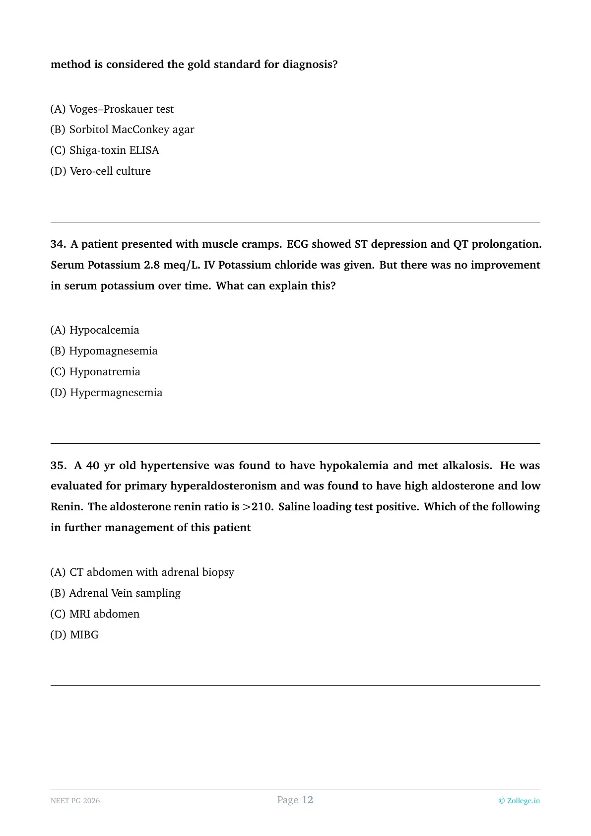
Several students from the same school develop severe abdominal cramps and bloody diarrhea. Shiga toxin-producing Escherichia coli (STEC/EHEC) is suspected. Which reference method is considered the gold standard for diagnosis?
View Solution
Step 1: Understanding the Question:
The scenario describes an outbreak of hemorrhagic colitis (bloody diarrhea) caused by Shiga Toxin-producing E. coli (STEC), such as O157:H7. We need to identify the "gold standard" or reference method for diagnosing the presence of the toxin or the organism's toxigenic potential.
Step 2: Key Formula or Approach:
While culture and ELISA are commonly used in clinical labs, "gold standard" usually refers to the definitive bioassay that proves the physiological effect of the toxin. Shiga toxin is also known as "Verotoxin."
Step 3: Detailed Explanation:
1. Vero-cell culture: This is the reference "gold standard" bioassay. Shiga toxins (Stx1 and Stx2) have a potent cytotoxic effect on Vero cells (a lineage of kidney epithelial cells from the African green monkey). The assay demonstrates the actual biological activity (cell death) caused by the toxin in the patient's stool sample or bacterial isolate.
2. Sorbitol MacConkey (SMAC) agar: This is the standard screening method. Most EHEC (O157:H7) do not ferment sorbitol and appear as colorless colonies. However, it is not the gold standard because it only identifies the phenotype, not the toxin production itself.
3. ELISA: This is a rapid test for the presence of the toxin proteins but is slightly less sensitive/specific than the cell culture bioassay or PCR for the stx genes.
Step 4: Final Answer:
The gold standard reference method for detecting the biological activity of Shiga toxin is the Vero-cell culture cytotoxicity assay.
A patient presented with muscle cramps. ECG showed ST depression and QT prolongation. Serum Potassium 2.8 meq/L. IV Potassium chloride was given. But there was no improvement in serum potassium over time. What can explain this?
View Solution
Step 1: Understanding the Question:
The patient has significant hypokalemia (K+ = 2.8) and symptoms (cramps, ECG changes). Despite receiving intravenous replacement (KCl), the potassium levels are not rising ("refractory hypokalemia"). We need to identify the underlying electrolyte deficiency that prevents potassium correction.
Step 2: Key Formula or Approach:
Magnesium and Potassium are physiologically linked in the renal tubules. Magnesium acts as a "gatekeeper" for potassium channels in the kidney.
Step 3: Detailed Explanation:
1. Role of Magnesium: Intracellular magnesium normally inhibits the ROMK (Renal Outer Medullary Potassium) channels in the thick ascending limb and collecting duct of the nephron.
2. The Defect: In a state of hypomagnesemia, this inhibition is lost. The ROMK channels remain wide open, allowing for the excessive and "uncontrolled" secretion of potassium into the urine.
3. Consequence: Because the kidney is constantly "leaking" potassium due to low magnesium, any potassium you provide intravenously is simply excreted in the urine. Therefore, the serum potassium level will not correct until the magnesium deficiency is fixed first.
4. ECG Findings: Both hypokalemia and hypomagnesemia cause similar ECG changes, including ST-segment depression, flattened T waves, and prominent U waves (which can lead to QT prolongation).
Step 4: Final Answer:
Hypomagnesemia is the classic cause of refractory hypokalemia that does not respond to potassium supplementation.
A 40 yr old hypertensive was found to have hypokalemia and met alkalosis. He was evaluated for primary hyperaldosteronism and was found to have high aldosterone and low Renin. The aldosterone renin ratio is $>$210. Saline loading test positive. Which of the following in further management of this patient
View Solution
Step 1: Understanding the Question:
The patient has the classic presentation of Primary Hyperaldosteronism (Conn's Syndrome): hypertension, hypokalemia, and metabolic alkalosis. The screening test (Aldosterone-Renin Ratio $>$ 20-30) is positive, and the confirmatory test (Saline Loading Test failing to suppress aldosterone) is also positive. We now need the next step in management.
Step 2: Key Formula or Approach:
The management algorithm for Primary Hyperaldosteronism is:
1. Screening (ARR).
2. Confirmation (Saline loading or Fludrocortisone suppression).
3. Lateralization (to distinguish between a unilateral adenoma, which is surgically curable, and bilateral hyperplasia, which is managed medically).
Step 3: Detailed Explanation:
1. Adrenal Vein Sampling (AVS): While a CT or MRI is usually the first imaging step to look for a mass, imaging alone is often misleading (it can miss small functional adenomas or find non-functional incidentalomas). AVS is the gold standard for lateralization. It involves sampling blood from both adrenal veins to compare aldosterone levels. If one side is significantly higher, it confirms a unilateral adenoma, and the patient is a candidate for adrenalectomy.
2. Biopsy: Adrenal biopsy (Option A) is contraindicated in the evaluation of functional adrenal tumors due to the risk of hypertensive crisis (if it's a pheochromocytoma) or tumor seeding.
3. MIBG: This is a nuclear scan used for identifying neuroendocrine tumors like Pheochromocytoma, not for hyperaldosteronism.
Step 4: Final Answer:
Following the confirmation of Primary Hyperaldosteronism, Adrenal Vein Sampling is the definitive next step to determine the site of hormone overproduction before considering surgery.
NEET PG 2026 Final Revision
*The article might have information for the previous academic years, please refer the official website of the exam.