
CBSE Class 12 Biology Question Paper 2025 (Set 3 - 57/4/3) with Solution Pdf


| Updated On - Feb 11, 2026
CBSE Class 12 Biology Question Paper with Solutions PDF is now available for download. CBSE conducted the Class 12 Biology examination on March 25, 2025. The question paper consists of 33 questions carrying a total of 70 marks. Section A includes 16 MCQs for 1 mark each, Section B contains 5 very short-answer questions for 2 marks each, Section C comprises 7 short-answer questions for 3 marks each, Section D comprises 2 Case-based questions carries 4 marks each and Section E comprises 3 long-answer questions carries 5 marks each.
CBSE Class 12 Biology Question Paper (Set 3 – 57/4/3) 2025 with Solution Pdf
| CBSE Class 12 Biology Question Paper | Download PDF | Check Solutions |
In its extended 'beads-on-string' form of chromatin, the 'string' in the 'beads-on-string' represent :
View Solution
Step 1: Understanding the Question:
The question asks to identify the component that represents the 'string' in the 'beads-on-string' model of chromatin structure.
Step 3: Detailed Explanation:
Chromatin is the complex of DNA and proteins found inside the nucleus of eukaryotic cells.
The fundamental repeating unit of chromatin is the nucleosome, which gives chromatin a 'beads-on-string' appearance under an electron microscope.
In this structure:
- The 'beads' are the nucleosomes. Each nucleosome consists of a core of eight histone proteins (a histone octamer) around which approximately 147 base pairs of DNA are wrapped.
- The 'string' is the stretch of DNA that connects two adjacent nucleosomes. This connecting DNA is called linker DNA.
Therefore, the 'string' in the 'beads-on-string' model represents the linker DNA.
Histones (B) form the 'beads'. NHC (Non-Histone Chromosomal) proteins (C) are other proteins involved in DNA compaction and regulation but are not the 'string'. 200 bp (D) refers to the approximate length of DNA in a nucleosome plus linker, not a protein component.
Step 4: Final Answer:
The correct answer is (A) Linker DNA, which connects the nucleosome 'beads'.
Quick Tip: Visualize the chromatin structure. The histones are like spools, and the DNA is the thread. The DNA wrapped around the spool is the bead (nucleosome), and the thread running between spools is the string (linker DNA). This analogy helps in remembering the components.
Given below are few statements with reference to oogenesis in a human female:
(i) An unequal division of the primary oocyte forms a large secondary oocyte and a tiny first polar body.
(ii) A couple of thousands of oogonia are formed within each fetal ovary during embryonic development.
(iii) Primary oocytes start meiosis-I but it gets arrested temporarily at prophase-I during fetal development.
(iv) Fertilisation induces the completion of the meiotic division of the secondary oocyte.
(v) A primary oocyte on meiosis forms an ovum and a polar body.
Choose the option with all true statements from the given options :
View Solution
Step 1: Understanding the Question:
The question requires us to evaluate five statements about oogenesis (the process of egg formation) in human females and identify the combination of statements that are all true.
Step 3: Detailed Explanation:
Let's analyze each statement:
- Statement (i): An unequal division of the primary oocyte forms a large secondary oocyte and a tiny first polar body. This is True. Meiosis I in the primary oocyte is a reductional division but results in unequal cytokinesis, conserving the cytoplasm for the secondary oocyte.
- Statement (ii): A couple of thousands of oogonia are formed within each fetal ovary during embryonic development. This is False. Millions (not thousands) of oogonia are formed during fetal development. No more oogonia are formed or added after birth.
- Statement (iii): Primary oocytes start meiosis-I but it gets arrested temporarily at prophase-I during fetal development. This is True. This arrested state continues until the female reaches puberty.
- Statement (iv): Fertilisation induces the completion of the meiotic division of the secondary oocyte. This is True. The secondary oocyte is arrested in metaphase-II. The entry of the sperm triggers the completion of meiosis-II, resulting in the formation of a mature ovum and a second polar body.
- Statement (v): A primary oocyte on meiosis forms an ovum and a polar body. This is False. A primary oocyte completes meiosis I to form a secondary oocyte and a first polar body. It does not directly form an ovum. The secondary oocyte forms the ovum after completing meiosis II.
Step 4: Final Answer:
The true statements are (i), (iii), and (iv). Therefore, option (D) is the correct choice.
Quick Tip: Remember the key arrest stages in oogenesis:
1. Prophase-I (arrest of primary oocyte, from fetal life to puberty).
2. Metaphase-II (arrest of secondary oocyte, from ovulation until fertilization).
This helps in quickly identifying correct and incorrect statements about the process.
Thalassemia and Sickle Cell Anaemia are both caused due to problem in globin molecule synthesis.
Select the correct statement :
View Solution
Step 1: Understanding the Question:
The question asks to differentiate between Thalassemia and Sickle Cell Anaemia, both of which are genetic disorders affecting globin synthesis. We need to identify the correct statement describing the nature of the defect in each.
Step 3: Detailed Explanation:
Let's differentiate between the two disorders:
- Thalassemia: This is a quantitative disorder. It is caused by a gene mutation that leads to a reduced rate of synthesis of one of the globin chains (\(\alpha\) or \(\beta\)) that make up haemoglobin. This results in the production of fewer globin molecules than normal, causing anaemia. So, statement (B) is correct.
- Sickle Cell Anaemia: This is a qualitative disorder. It is caused by a point mutation in the \(\beta\)-globin gene, which leads to the substitution of Glutamic acid by Valine at the sixth position of the \(\beta\)-globin chain. This results in the synthesis of an abnormal haemoglobin molecule (HbS), which functions incorrectly, causing red blood cells to become sickle-shaped under low oxygen tension. The quantity of globin produced is normal, but its quality is defective.
Now let's evaluate the options:
- (A) Both are due to a quantitative defect. This is false; Sickle cell anaemia is qualitative.
- (B) Thalassemia is due to less synthesis of globin molecules. This is true, as it's a quantitative defect.
- (C) Sickle cell anaemia is due to a quantitative problem. This is false; it's a qualitative problem.
- (D) Both are due to qualitative defect. This is false; Thalassemia is quantitative.
Step 4: Final Answer:
The only correct statement is (B). Thalassemia is a quantitative problem characterized by the reduced synthesis of globin molecules.
Quick Tip: Remember:
- Thalassemia = \textbf{Quan}titative defect (less quantity of globin).
- Sickle Cell Anaemia = \textbf{Qual}itative defect (poor quality of globin).
This simple association helps to avoid confusion between these two common genetic disorders.
A pedigree chart in shown below for a disease that is autosomal dominant. The genetic make-up of the first generation is :

View Solution
Step 1: Understanding the Question:
The question provides a pedigree chart for an autosomal dominant disease and asks for the genotypes of the parents in the first generation (Generation I).
In a pedigree chart:
- A circle represents a female.
- A square represents a male.
- A shaded symbol represents an affected individual.
- An unshaded symbol represents an unaffected individual.
Step 3: Detailed Explanation:
Analyzing Generation I:
- There is one affected individual (shaded circle, a female) and one unaffected individual (unshaded square, a male).
Determining Genotypes:
- The disease is autosomal dominant. Let 'A' be the dominant allele causing the disease, and 'a' be the recessive allele for the normal phenotype.
- An unaffected individual must have a homozygous recessive genotype, which is aa. Therefore, the male in Generation I has the genotype aa.
- An affected individual must have at least one dominant allele, so their genotype is either AA or Aa.
Using Offspring Information (Generation II):
- The couple in Generation I has offspring in Generation II. We can see that they have both affected and unaffected children.
- They have an unaffected daughter (unshaded circle) and an unaffected son (unshaded square).
- For an offspring to be unaffected (genotype aa), they must inherit one 'a' allele from each parent.
- We already know the father is 'aa', so he can only pass on an 'a' allele.
- The affected mother must also be able to pass on an 'a' allele to her unaffected children. This means her genotype cannot be AA.
- Therefore, the affected mother must be heterozygous, with the genotype Aa.
Step 4: Final Answer:
The genetic make-up of the first generation is: Affected female (Aa) and Unaffected male (aa). This corresponds to option (B).
Quick Tip: For dominant traits in a pedigree, look for affected parents having an unaffected child. This immediately tells you that the affected parent(s) must be heterozygous. An unaffected child (aa) can only be born if both parents can contribute an 'a' allele.
Allergy in humans is a result of release of the following chemicals from the mast cells :
View Solution
Step 1: Understanding the Question:
The question asks to identify the chemicals that are released from mast cells during an allergic reaction in humans.
Step 3: Detailed Explanation:
An allergy is an exaggerated or hypersensitive response of the immune system to certain antigens present in the environment, known as allergens.
When a person is exposed to an allergen they are sensitive to, their body produces IgE antibodies. These IgE antibodies attach to the surface of mast cells and basophils.
Upon subsequent exposure to the same allergen, the allergen binds to the IgE antibodies on the mast cells. This triggers the mast cells to degranulate, which means they release potent inflammatory chemicals stored in their granules into the surrounding tissues.
The primary chemicals released by mast cells during an allergic reaction are histamine and serotonin. These chemicals cause the symptoms associated with allergies, such as vasodilation (widening of blood vessels), increased permeability of capillaries, contraction of smooth muscles (e.g., in the bronchi), and increased mucus secretion.
The other options are neurotransmitters or hormones with different primary functions:
- (B) Adrenaline (hormone) and Acetylcholine (neurotransmitter).
- (C) Dopamine and Nor-adrenalin (neurotransmitters).
- (D) Endorphins and GABA (neurotransmitters).
Step 4: Final Answer:
The correct chemicals released from mast cells during an allergic reaction are Histamine and Serotonin. Thus, option (A) is correct.
Quick Tip: Associate allergies with "histamine." This is why antihistamine medications (like Benadryl or Claritin) are used to treat allergy symptoms. They work by blocking the effects of histamine released by mast cells.
Select the statements that are true for outbreeding devices in the flowering plants.
(i) They discourage cross-pollination and ensure self-pollination.
(ii) They are advantageous as they lead to inbreeding depression.
(iii) Self-incompatibility prevents self-pollen from fertilising the ovules.
(iv) To prevent self-pollination in some species pollen release and stigma receptivity are not synchronised.
Choose the correct answer :
View Solution
Step 1: Understanding the Question:
The question asks to identify the true statements about outbreeding devices in flowering plants. Outbreeding devices are mechanisms that plants have evolved to encourage cross-pollination and discourage self-pollination.
Step 3: Detailed Explanation:
Let's analyze each statement:
- Statement (i): They discourage cross-pollination and ensure self-pollination. This is False. Outbreeding devices do the exact opposite; they encourage cross-pollination (xenogamy) and discourage self-pollination (autogamy).
- Statement (ii): They are advantageous as they lead to inbreeding depression. This is False. They are advantageous because they prevent inbreeding depression. Inbreeding depression is the reduced biological fitness in a population as a result of continuous self-pollination or breeding of related individuals.
- Statement (iii): Self-incompatibility prevents self-pollen from fertilising the ovules. This is True. Self-incompatibility is a genetic mechanism that prevents self-pollen (from the same flower or another flower on the same plant) from germinating on the stigma or from the pollen tube growing through the style, thus inhibiting fertilization. It is a key outbreeding device.
- Statement (iv): To prevent self-pollination in some species pollen release and stigma receptivity are not synchronised. This is True. This mechanism is called dichogamy. If the pollen is released before the stigma becomes receptive (protandry) or if the stigma becomes receptive before the pollen is released (protogyny), self-pollination is prevented.
Step 4: Final Answer:
The true statements are (iii) and (iv). Therefore, the correct option is (C).
Quick Tip: Remember that "outbreeding" means breeding with others (cross-pollination). Therefore, any device or mechanism described must logically support this goal. This helps to quickly eliminate options that suggest promoting self-pollination.
The first human-like homonid evolved during human evolution is supposed to be :
View Solution
Step 1: Understanding the Question:
The question asks to identify the species considered to be the first "human-like" hominid in the course of human evolution.
Step 3: Detailed Explanation:
Let's look at the evolutionary sequence of the hominids listed:
1. Australopithecines (pre-Homo): These were early hominids who were bipedal but had smaller brains. They are considered ancestors to the genus *Homo*.
2. Homo habilis ('Handy man'): This species is considered the earliest member of the genus *Homo*. They lived about 2.4 to 1.4 million years ago. Their brain was larger than that of the Australopithecines (about 650-800 cc), and they are associated with the first stone tools. Their more human-like features and tool-making ability lead to them being called the first "human-like" hominid.
3. Homo erectus ('Upright man'): Evolved after *Homo habilis*, around 1.8 million years ago. They had a larger brain (about 900 cc), were more proficient toolmakers, and likely used fire.
4. Neanderthal man (Homo neanderthalensis): A much later species (or subspecies) that lived in Europe and Asia from about 400,000 to 40,000 years ago. They had brains as large as or larger than modern humans.
5. Homo sapiens ('Wise man'): This is the species of modern humans, which appeared in Africa around 300,000 years ago.
Based on this timeline, *Homo habilis* is the first species in the genus *Homo* and is thus regarded as the first human-like hominid.
Step 4: Final Answer:
The correct answer is (B) Homo habilis.
Quick Tip: Remember the general order of human evolution: *Australopithecus* -> *Homo habilis* -> *Homo erectus* -> *Homo neanderthalensis* -> *Homo sapiens*. *Habilis* means "handy" or "skilful," referring to their tool use, which is a key human-like trait.
Use the given information to select the amino acid attached to the 3' end of tRNA during the process of translation, if the coding strand of the structural gene being transcribed has the nucleotide sequence ‘TCC’.
Codons for the amino acids :
AGG – Arginine
AAG – Lysine
UCC – Serine
GGA – Glycine
View Solution
Step 1: Understanding the Question:
The question asks us to find the amino acid that will be brought by a tRNA during translation, given the sequence of the coding strand of the DNA.
Step 2: Key Formula or Approach:
The process involves two main steps: Transcription (DNA to mRNA) and Translation (mRNA to protein).
1. Determine the mRNA codon from the DNA coding strand.
2. Use the provided table to match the mRNA codon to its corresponding amino acid.
Step 3: Detailed Explanation:
Part 1: Transcription
We are given the DNA coding strand sequence: 5'-TCC-3'.
There are two ways to find the mRNA sequence:
- Method 1 (using the template strand): The template strand is complementary to the coding strand. So, the template strand is 3'-AGG-5'. The mRNA is transcribed from the template strand and will be complementary to it (with U instead of T). So, the mRNA codon is 5'-UCC-3'.
- Method 2 (direct from coding strand): The mRNA sequence is identical to the DNA coding strand, with the only difference being that Thymine (T) is replaced by Uracil (U). So, we can directly replace T with U in the coding strand sequence.
DNA coding strand: TCC
mRNA codon: UCC
Part 2: Translation
The mRNA codon is UCC.
Now, we look at the provided table of codons for amino acids:
- AGG – Arginine
- AAG – Lysine
- UCC – Serine
- GGA – Glycine
The codon UCC codes for the amino acid Serine. This is the amino acid that will be attached to the corresponding tRNA.
Step 4: Final Answer:
The amino acid corresponding to the DNA coding strand 'TCC' is Serine. Therefore, option (C) is correct.
Quick Tip: The key here is the term "coding strand." The mRNA sequence will be the same as the coding strand, just with U replacing T. This is a shortcut that saves you the step of first finding the template strand.
The most widely used transgenic animal for testing the safety of vaccine before they are used in humans is :
View Solution
Step 1: Understanding the Question:
The question asks to identify the most common transgenic animal model used for vaccine safety testing prior to human trials.
Step 3: Detailed Explanation:
Transgenic animals are animals that have had a foreign gene deliberately inserted into their genome. They are widely used in biomedical research to study diseases and test the safety and efficacy of new drugs and vaccines.
Among the options provided:
- Mice are overwhelmingly the most common animal model in biomedical research, including for vaccine safety. Over 95% of all lab animals are mice and rats.
Reasons for the widespread use of mice include:
- Their physiological and genetic similarity to humans.
- Short lifespan and rapid reproduction rate, which allows for studying effects across generations quickly.
- Small size, making them easy to house and handle.
- Well-understood genetics and the availability of many established inbred and transgenic strains.
- Rabbits, pigs, and sheep are also used in research, but to a much lesser extent than mice, especially for initial large-scale safety and efficacy testing of vaccines. For example, transgenic pigs are used for organ transplant research, and sheep for studying certain genetic diseases.
Step 4: Final Answer:
Given their prevalence in biomedical research, mice are the most widely used transgenic animals for testing vaccine safety. The correct answer is (D).
Quick Tip: When asked about the "most common" or "most widely used" animal in general biomedical research or genetic studies, the answer is almost always mice. Their convenience and genetic tractability make them the workhorse of modern biology.
To prove that DNA replication is semiconservative, the diagram that correctly represents the DNA extracted from the culture after 40 minutes of centrifugation in the experiment by Matthew Meselson and Franklin Stahl is :
- (A)
![]()
- (B)
![]()
- (C)
![]()
- (D)
![]()


View Solution
Step 1: Understanding the Question:
The question refers to the Meselson-Stahl experiment, which demonstrated the semiconservative nature of DNA replication. We need to identify the correct diagram showing the result of CsCl density gradient centrifugation after two generations (40 minutes, assuming a 20-minute generation time for E. coli).
Step 3: Detailed Explanation:
Initial State (Generation 0):
- E. coli bacteria are grown for many generations in a medium containing a heavy isotope of nitrogen, \(^{15}\)N.
- As a result, all of their DNA is heavy (\(^{15}\)N/\(^{15}\)N). This would form a single, low band in the centrifuge tube.
After 20 minutes (Generation 1):
- The bacteria are transferred to a medium containing the normal, lighter isotope, \(^{14}\)N.
- After one round of replication (20 minutes), each new DNA molecule consists of one old \(^{15}\)N strand and one new \(^{14}\)N strand.
- This results in hybrid DNA (\(^{15}\)N/\(^{14}\)N) of intermediate density. It would form a single band positioned between the heavy \(^{15}\)N and light \(^{14}\)N positions. This matches diagram (C).
After 40 minutes (Generation 2):
- The bacteria undergo a second round of replication in the \(^{14}\)N medium.
- The two hybrid DNA molecules from Generation 1 unwind and replicate.
- The \(^{15}\)N strand of each hybrid pairs with a new \(^{14}\)N strand, forming a hybrid molecule (\(^{15}\)N/\(^{14}\)N).
- The \(^{14}\)N strand of each hybrid pairs with a new \(^{14}\)N strand, forming a light molecule (\(^{14}\)N/\(^{14}\)N).
- This results in a population of DNA molecules where 50% are hybrid (\(^{15}\)N/\(^{14}\)N) and 50% are light (\(^{14}\)N/\(^{14}\)N).
- In the centrifuge tube, this will appear as two distinct bands of equal intensity: one at the intermediate position and one at the light (higher) position.
Analyzing the Diagrams:
- Diagram (A) shows two bands: one intermediate and one light. This correctly represents the result after 40 minutes.
- Diagram (B) shows a heavy and a light band, which would be the result of conservative replication.
- Diagram (C) shows a single intermediate band, which is the result after 20 minutes.
- Diagram (D) is incorrect.
Step 4: Final Answer:
The correct representation after 40 minutes (two generations) is two equal bands, one hybrid and one light. This is shown in diagram (A).
Quick Tip: Remember the progression in the Meselson-Stahl experiment:
- Gen 0: 1 Heavy band.
- Gen 1: 1 Intermediate band.
- Gen 2: 1 Intermediate band + 1 Light band (equal amounts).
- Gen 3: 1 Intermediate band + 1 larger Light band.
This pattern is the classic proof of semiconservative replication.
Statins, a bioactive molecule used for human welfare, are sourced from :
View Solution
Step 1: Understanding the Question:
The question asks for the microbial source of statins, which are bioactive molecules used as medicine.
Step 3: Detailed Explanation:
Statins are a class of drugs used to lower cholesterol levels in the blood. They act by competitively inhibiting the enzyme HMG-CoA reductase, which plays a central role in the production of cholesterol in the liver.
Let's examine the sources listed in the options:
- (A) Trichoderma polysporum: This fungus is the source of cyclosporin A, an immunosuppressive agent used in organ transplant patients.
- (B) Monascus purpureus: This is a species of yeast that is used to produce statins. The statins it produces are used as blood-cholesterol lowering agents.
- (C) Propionibacterium sharmanii: This bacterium is used in the production of Swiss cheese, where it is responsible for the large holes and characteristic flavour due to the production of CO\(_2\). It is also a source of Vitamin B12.
- (D) Aspergillus niger: This fungus is used for the industrial production of citric acid.
Step 4: Final Answer:
The correct source for statins is the yeast *Monascus purpureus*. Therefore, option (B) is correct.
Quick Tip: Create a table to remember important microbes and their products:
- *\textbf{Monascus purpureus}* \(\rightarrow{}\) Statins (cholesterol lowering).
- *\textbf{Trichoderma polysporum}* \(\rightarrow{}\) Cyclosporin A (immunosuppressant).
- *\textbf{Streptococcus}* \(\rightarrow{}\) Streptokinase (clot buster).
- *\textbf{Aspergillus niger}* \(\rightarrow{}\) Citric acid.
This helps in quick recall for matching-type questions.
In a pea plant (Pisum sativum), green pod colour is dominant over yellow pod colour. The phenotypic ratio of the offspring in the first generation of a cross in which both parents are heterozygous for green pod colour will be :
View Solution
Step 1: Understanding the Question:
The question describes a classic Mendelian monohybrid cross. We need to find the phenotypic ratio of the offspring from a cross between two pea plants that are both heterozygous for pod colour. We are told that green pod colour is dominant.
Step 2: Key Formula or Approach:
The approach is to set up a Punnett square for a monohybrid cross (Gg x Gg).
- Define the alleles.
- Determine the genotypes of the parents.
- Set up the Punnett square to find the genotypes of the offspring.
- Determine the phenotypes based on the genotypes and the dominance relationship.
- Calculate the phenotypic ratio.
Step 3: Detailed Explanation:
1. Define Alleles:
- Let 'G' represent the dominant allele for green pod colour.
- Let 'g' represent the recessive allele for yellow pod colour.
2. Parental Genotypes:
- Both parents are heterozygous for green pod colour. This means their genotype is Gg.
3. The Cross:
- The cross is Gg x Gg.
4. Punnett Square:
The possible gametes from each parent are G and g.
\begin{tabular{c|c|c|
\multicolumn{1{c{ & \multicolumn{1{c{G & \multicolumn{1{c{g
\cline{2-3
G & GG & Gg
\cline{2-3
g & Gg & gg
\cline{2-3
\end{tabular
5. Offspring Genotypes and Phenotypes:
From the Punnett square, the genotypic ratio of the offspring is 1 GG : 2 Gg : 1 gg.
- GG: Homozygous dominant, phenotype is Green pods.
- Gg: Heterozygous, phenotype is Green pods (since G is dominant).
- gg: Homozygous recessive, phenotype is Yellow pods.
6. Phenotypic Ratio:
- Number of offspring with green pods = 1 (GG) + 2 (Gg) = 3
- Number of offspring with yellow pods = 1 (gg)
- The phenotypic ratio of green pods to yellow pods is 3:1.
Step 4: Final Answer:
The phenotypic ratio for a heterozygous monohybrid cross is 3 (dominant phenotype) : 1 (recessive phenotype). The correct option is (D) 3:1.
Quick Tip: For any simple Mendelian monohybrid cross between two heterozygous parents (e.g., Tt x Tt, Gg x Gg), the expected ratios are always:
- Genotypic ratio: 1 : 2 : 1 (Homozygous Dominant : Heterozygous : Homozygous Recessive).
- Phenotypic ratio: 3 : 1 (Dominant Phenotype : Recessive Phenotype).
Memorizing these standard ratios can save a lot of time in exams.
Assertion (A) : ABO blood grouping in humans is an example of multiple allelism.
Reason (R) : More than two genes in a population govern the same character in ABO blood grouping in humans.
View Solution
Step 1: Understanding the Question:
This question requires an analysis of an assertion and a reason related to the genetics of ABO blood grouping in humans. We must determine if each statement is true and if the reason correctly explains the assertion.
Step 3: Detailed Explanation:
Analysis of Assertion (A):
The assertion states that ABO blood grouping is an example of multiple allelism. Multiple allelism is a condition where more than two alleles for a particular gene exist within a population. The ABO blood group system is controlled by the gene 'I', which has three alleles: I\(^A\), I\(^B\), and i. Since there are three alleles for this single gene, it is a classic example of multiple allelism. Thus, Assertion (A) is true.
Analysis of Reason (R):
The reason states that "More than two genes in a population govern the same character". This statement is incorrect. The ABO blood group is governed by a single gene (the 'I' gene) which has \textit{multiple alleles (I\(^A\), I\(^B\), i). The phenomenon where multiple genes control a single character is called polygenic inheritance, not multiple allelism. Therefore, Reason (R) is false.
Step 4: Final Answer:
Assertion (A) is true because the ABO system is controlled by three alleles of a single gene. Reason (R) is false because it incorrectly describes polygenic inheritance instead of multiple allelism. Thus, the correct option is (C).
Quick Tip: Distinguish clearly between these genetic terms:
- \textbf{Multiple Alleles: One gene, more than two alleles (e.g., ABO blood groups).
- \textbf{Polygenic Inheritance:} One trait, controlled by multiple genes (e.g., human skin colour, height).
This distinction is crucial for correctly answering assertion-reason questions in genetics.
Assertion (A) : The meristems are grown ‘in vitro’ to obtain virus-free plants from an infected plant.
Reason (R) : If the plant is infected with a virus, the roots and the stems are free of virus.
View Solution
Step 1: Understanding the Question:
The question presents an assertion and a reason related to plant tissue culture for obtaining virus-free plants. We need to evaluate the truthfulness of both statements and their relationship.
Step 3: Detailed Explanation:
Analysis of Assertion (A):
The assertion states that meristems are cultured in vitro to get virus-free plants. This is a standard and true technique in plant biotechnology called meristem culture. The reason meristems (apical and axillary) are used is that the rate of cell division in the meristematic tissue is very high, and it often outpaces the rate of viral multiplication and movement. As a result, the meristematic tip is usually free of viruses even if the rest of the plant is infected. Thus, Assertion (A) is true.
Analysis of Reason (R):
The reason states that if a plant is infected with a virus, the roots and stems are free of the virus. This is generally false. Viruses are systemic pathogens, meaning they typically spread throughout the plant's vascular system (phloem and xylem), infecting various parts, including the stems, leaves, and roots. Only the meristematic tips are reliably virus-free. Thus, Reason (R) is false.
Step 4: Final Answer:
Assertion (A) is true because meristem culture is a valid method to obtain virus-free plants. Reason (R) is false because viruses usually infect most parts of the plant, not just specific organs. Therefore, the correct option is (C).
Quick Tip: Remember that the key to virus-free plant propagation lies in the unique property of the meristem. Its rapid cell division rate is the reason it remains uninfected. Don't confuse this with other plant parts like mature stems or roots, which are typically susceptible to systemic viral infections.
Assertion (A) : A person infected with malaria suffers from chill and high fever, recurring every three or four days.
Reason (R) : The parasite attacks the RBC resulting in their rupture and release of haemozoin.
View Solution
Step 1: Understanding the Question:
The question asks to evaluate an assertion about the symptoms of malaria and a reason explaining the physiological cause of these symptoms.
Step 3: Detailed Explanation:
Analysis of Assertion (A):
The assertion states that a malaria-infected person experiences recurring chills and high fever every three to four days. This is the classic clinical symptom of malaria. The malarial parasite, *Plasmodium*, has a life cycle in humans that involves the asexual reproduction within red blood cells (RBCs). The cycle of RBC rupture and release of new parasites (merozoites) occurs in a synchronized manner, leading to these periodic episodes of fever and chills. The length of the cycle (e.g., 48 or 72 hours) depends on the *Plasmodium* species. So, Assertion (A) is true.
Analysis of Reason (R):
The reason states that the parasite attacks RBCs, causing their rupture and the release of a substance called haemozoin. This is also true. Inside the RBC, the parasite feeds on haemoglobin. The toxic heme part of haemoglobin is converted into an insoluble crystalline substance called haemozoin. When the infected RBCs rupture to release the next generation of parasites, this haemozoin is also released into the bloodstream. Haemozoin is a toxic substance that triggers the immune response, leading to the characteristic chills and high fever. Thus, Reason (R) is true.
Relationship between A and R:
The release of haemozoin upon the rupture of RBCs is the direct cause of the chills and fever mentioned in the assertion. Therefore, the reason correctly explains the assertion.
Step 4: Final Answer:
Both the assertion and the reason are true, and the reason provides the correct explanation for the assertion. Therefore, option (A) is the correct answer.
Quick Tip: Associate the symptoms of malaria (chills and fever) directly with the release of the toxin \textbf{haemozoin}. This release happens when the red blood cells burst. Remembering this direct causal link helps in answering questions about the pathogenesis of malaria.
Assertion (A): ‘Saheli', an oral contraceptive inhibits ovulation and increases phagocytosis of sperms.
Reason (R) : It is a non-steroidal preparation.
View Solution
Step 1: Understanding the Question:
The question asks to evaluate an assertion about the mechanism of action of the contraceptive pill 'Saheli' and a reason describing its chemical nature.
Step 3: Detailed Explanation:
Analysis of Assertion (A):
The assertion states that 'Saheli' inhibits ovulation and increases phagocytosis of sperms. This is incorrect. 'Saheli' (Centchroman) is a selective estrogen receptor modulator (SERM). Its primary mechanism of action is to prevent the implantation of the fertilised ovum in the uterus. It does not consistently inhibit ovulation, nor does it increase the phagocytosis of sperms. Traditional combined oral contraceptive pills (containing estrogen and progestin) work by inhibiting ovulation. Thus, Assertion (A) is false.
Analysis of Reason (R):
The reason states that 'Saheli' is a non-steroidal preparation. This is true. 'Saheli' was developed in India by the Central Drug Research Institute (CDRI) and is notable for being one of the first non-steroidal oral contraceptives. Its non-steroidal nature results in fewer side effects compared to traditional steroidal pills. Thus, Reason (R) is true.
Step 4: Final Answer:
Assertion (A) is false because it misrepresents the mechanism of action of 'Saheli'. Reason (R) is true because 'Saheli' is indeed a non-steroidal drug. Therefore, the correct option is (D).
Quick Tip: Remember the key difference between 'Saheli' and other common birth control pills. Most pills are steroidal and prevent ovulation. 'Saheli' is \textbf{non-steroidal} and works primarily by preventing \textbf{implantation}. This distinction is frequently tested.
Question 17:
The basic scheme of the essential steps involved in the process of recombinant DNA technology is summarized below in the form of a flow diagram. Study the given flow diagram and answer the questions that follow.
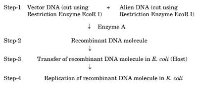
(a). Name the enzyme used in Step-1 to join the cut plasmid and alien DNA.
View Solution
Step 1: Understanding the Question:
The question refers to a flowchart of recombinant DNA technology and asks to identify "Enzyme A," which is used to join the vector DNA and the alien DNA after they have both been cut.
Step 3: Detailed Explanation:
In the process of creating a recombinant DNA molecule, after both the vector (like a plasmid) and the foreign DNA have been cut with the same restriction enzyme, their complementary "sticky ends" anneal (pair up). However, this pairing is temporary and held by weak hydrogen bonds. To create a stable recombinant molecule, the sugar-phosphate backbones of the DNA strands must be joined. The enzyme that catalyzes the formation of these phosphodiester bonds, effectively "pasting" or "ligating" the DNA fragments together, is DNA Ligase.
Quick Tip: Think of recombinant DNA technology as a "cut and paste" process. Restriction enzymes are the "molecular scissors" that cut, and DNA Ligase is the "molecular glue" that pastes the pieces together.
State the technical term used for Step-3.
View Solution
Step 1: Understanding the Question:
The question asks for the technical name of Step-3 in the provided flowchart, which describes the "Transfer of recombinant DNA molecule in E. coli (Host)".
Step 3: Detailed Explanation:
The process of introducing a piece of foreign DNA (in this case, the recombinant plasmid) into a host bacterium is known as Transformation. For this to occur, the host bacterial cells must be made "competent" to take up the DNA from the surrounding environment. This is often achieved by treating the cells with a specific concentration of a divalent cation, such as calcium, followed by a heat shock.
Quick Tip: Remember that transformation involves making a cell take up naked DNA from its environment. Other methods of gene transfer include transduction (via virus) and conjugation (via cell-to-cell contact).
Justify the use of same Restriction Enzyme EcoR I to cut both the vector DNA and the alien DNA.
View Solution
Step 1: Understanding the Question:
The question asks for the reason why the same restriction enzyme must be used to cleave both the gene of interest (alien DNA) and the vehicle for carrying it (vector DNA).
Step 3: Detailed Explanation:
Restriction enzymes are highly specific, recognizing and cutting at particular palindromic DNA sequences. Many, like EcoR I, make staggered cuts, which produce short, single-stranded overhangs. These overhangs are called "sticky ends" because they have a tendency to form hydrogen bonds with complementary sequences.
By using the same enzyme (EcoR I) on both DNA sources:
The vector DNA is cut, creating a specific sticky end.
The alien DNA is also cut, creating the exact same complementary sticky end.
This ensures that the "end" of the alien DNA fragment will perfectly match and anneal to the "end" of the opened vector. If different restriction enzymes were used, the sticky ends would not be complementary and would not be able to join, preventing the formation of a recombinant DNA molecule.
Quick Tip: Imagine you have a puzzle piece and a puzzle board. You must use the same-shaped cutter on both the piece and the hole in the board for them to fit together. Using different restriction enzymes is like using a star-shaped cutter for the piece and a circle-shaped cutter for the hole—they won't fit!
Explain how the interaction between sea anemone and clownfish is one of the best examples of commensalism in nature.
View Solution
Step 1: Understanding the Question:
The question asks for an explanation of why the relationship between a sea anemone and a clownfish is a prime example of commensalism.
Step 3: Detailed Explanation:
1. Definition of Commensalism:
Commensalism is a type of symbiotic relationship between two different species in which one species benefits, and the other species is neither harmed nor benefited (+/0 interaction).
2. Analysis of the Interaction:
- Benefit to the Clownfish (+): The sea anemone has stinging tentacles (containing nematocysts) that it uses to capture prey and deter predators. The clownfish has a protective mucus coating on its skin that makes it immune to the anemone's stings. By living within the tentacles, the clownfish gains significant protection from its own predators, which cannot tolerate the anemone's stings.
- Effect on the Sea Anemone (0): In the classic definition of this interaction, the sea anemone is not significantly affected by the clownfish's presence. The clownfish does not harm the anemone, nor does it provide a clear, substantial benefit. It simply uses the anemone for shelter.
3. Conclusion:
Since the clownfish clearly benefits (gains protection) and the sea anemone is largely unaffected, their relationship perfectly fits the +/0 pattern of commensalism.
Quick Tip: Remember the three main types of symbiosis by their interaction signs:
- \textbf{Mutualism (+/+):} Both benefit (e.g., lichens - algae and fungus).
- \textbf{Commensalism (+/0):} One benefits, one is unaffected (e.g., clownfish and anemone).
- \textbf{Parasitism (+/-):} One benefits, one is harmed (e.g., tapeworm and human).
OR
Question 18 (b):
Correctly depict (also indicate the trophic level) and describe the ecological pyramid of biomass in sea with 40 standing crop of phytoplankton supporting 90 standing crop of zooplankton which further supports 120 small fishes.
- \textbf{Trophic Level 1 (Producers):} Phytoplankton (Standing crop = 40 units)
- \textbf{Trophic Level 2 (Primary Consumers):} Zooplankton (Standing crop = 90 units)
- \textbf{Trophic Level 3 (Secondary Consumers):} Small fishes (Standing crop = 120 units)
\textbf{Description:} The pyramid is inverted because the biomass of the producers (phytoplankton) at any given time is less than the biomass of the consumers (zooplankton and fish) that it supports. This is possible due to the very high turnover rate (rapid reproduction and short lifespan) of phytoplankton.
View Solution
Step 1: Understanding the Question:
The question asks to depict and describe the pyramid of biomass for a specific marine food chain, given the standing crop values for each trophic level.
Step 3: Detailed Explanation:
1. Identifying Trophic Levels:
- The base of the food chain is phytoplankton, which are producers. Trophic Level 1 (T1). Standing crop = 40.
- Zooplankton feed on phytoplankton, so they are primary consumers. Trophic Level 2 (T2). Standing crop = 90.
- Small fishes feed on zooplankton, so they are secondary consumers. Trophic Level 3 (T3). Standing crop = 120.
2. Depicting the Pyramid of Biomass:
An ecological pyramid represents the amount of biomass at different trophic levels. The base is always the producer level (T1).
- Base (T1): Phytoplankton = 40
- Middle (T2): Zooplankton = 90
- Top (T3): Small fishes = 120
Since the biomass increases at each successive trophic level (40 < 90 < 120), the pyramid will be inverted. It will have a narrow base, a wider middle section, and the widest top section.
3. Describing the Inverted Pyramid:
The pyramid of biomass in this marine ecosystem is inverted. This is a characteristic feature of many aquatic ecosystems. The reason for this apparent paradox is the concept of "standing crop" versus "productivity." Standing crop is the biomass present at a particular moment. Phytoplankton, despite having a small standing crop, are extremely productive. They have a very short lifespan and reproduce very rapidly. Therefore, a small biomass of phytoplankton can support a much larger biomass of zooplankton because the phytoplankton are consumed and replaced at a very high rate. The zooplankton, having a longer lifespan, accumulate biomass over time. The high turnover rate of the producers is key to sustaining the larger consumer biomass.
Quick Tip: Remember that while the pyramid of \textbf{energy} is always upright, the pyramid of \textbf{biomass} can be inverted. The classic example of an inverted pyramid of biomass is a pond or ocean ecosystem, due to the high turnover rate of phytoplankton.
Explain how the immunity of a person is affected if there is atrophy (degeneration) of the thymus gland at an early stage of life.
View Solution
Step 1: Understanding the Question:
The question asks about the immunological consequences of the early degeneration of the thymus gland.
Step 3: Detailed Explanation:
1. Role of the Thymus Gland:
The thymus is a primary lymphoid organ whose main function is to serve as the site for the maturation and differentiation of T-lymphocytes (T-cells).
2. Consequence of Early Thymus Atrophy:
If the thymus gland degenerates at an early stage of life, the maturation process of T-cells will be severely impaired. This leads to:
- A drastic reduction in the number of functional T-cells.
- A collapse of cell-mediated immunity (CMI), leaving the person vulnerable to viral infections, fungal infections, and certain cancers.
- A significant weakening of humoral immunity, as the lack of helper T-cells would prevent B-cells from mounting an effective antibody response.
In summary, the person's entire adaptive immune system would be crippled, making them highly susceptible to recurrent and life-threatening infections.
Quick Tip: Remember the "T" in T-cells stands for Thymus. No Thymus, no functional T-cells. No T-cells means a failure of both cell-mediated immunity and a severely hampered antibody response.
OR
Question 19 (b):
(i) What are interferons ? Explain their role in providing immunity to a person.
(ii) Which category of innate immunity defence barrier can interferons be classified into ?
\textbf{(i)} Interferons are signaling proteins (cytokines) produced by virus-infected cells. They do not save the infected cell, but they are released to signal nearby uninfected cells to produce antiviral proteins. This protects the neighboring cells from viral infection and helps to limit the spread of the virus.
\textbf{(ii)} Interferons are classified under the \textbf{cytokine barrier} of innate immunity.
View Solution
Step 1: Understanding the Question:
The question asks for the definition and function of interferons, and their classification within the innate immune system.
Step 3: Detailed Explanation:
(i) Definition and Role of Interferons:
Interferons (IFNs) are proteins named for their ability to "interfere" with viral replication.
- Mechanism: When a cell is infected by a virus, it produces and releases interferons. These interferons travel to adjacent, uninfected cells and bind to their surface receptors. This signal induces the uninfected cells to produce a range of antiviral proteins, creating an "antiviral state" which blocks viral replication if the virus tries to enter these cells.
- Role in Immunity: They act as a crucial part of the early, non-specific (innate) immune response to viral infections, acting as a local alarm system to contain the virus.
(ii) Classification in Innate Immunity:
Innate immunity consists of four types of barriers. Interferons fall under the fourth category:
1. Physical barriers (Skin)
2. Physiological barriers (Stomach acid)
3. Cellular barriers (Phagocytes)
4. Cytokine barriers (Interferons)
Quick Tip: Think of interferons as a "warning signal" sent by a virus-infected cell. The signal doesn't help the cell that sent it, but it warns its neighbors to "lock their doors" (by producing antiviral proteins) before the virus can get to them.
Assume that the given mRNA (start site is not depicted) is theoretically translated in two reading frames.
(a) Translation starting from the first nucleotide (Reading frame 1)
(b) Translation starting from the second nucleotide (Reading frame 2)

Answer the following question
How many amino acids will be specified in case (a) and case (b) on translation ? Justify your answer.
\textbf{Case (a):} 7 amino acids.
\textbf{Case (b):} 5 amino acids.
\textbf{Justification:} In Frame 1, the 22-nucleotide sequence allows for 7 complete codons, specifying 7 amino acids. In Frame 2, the sequence also has 7 complete codons, but the 6th codon (UAA) is a stop codon. Translation terminates at a stop codon, so only the 5 codons preceding it are translated into a polypeptide chain of 5 amino acids.
View Solution
Step 1: Understanding the Question:
The question asks for the number of amino acids coded by two mRNA sequences, considering that translation reads the code in non-overlapping triplets and stops at a stop codon.
Step 3: Detailed Explanation:
Case (a) - Reading Frame 1:
- Sequence: 5' – CUCGCUUGCCGAUCAAGGGUUA – 3' (22 nt)
- Grouping into codons from the start: CUC | GCU | UGC | CGA | UCA | AGG | GUU | A
- There are 7 complete codons. Assuming none are stop codons, 7 amino acids will be specified.
Case (b) - Reading Frame 2:
- Sequence: 5' – GUGGCACUCAGUCCUUAAUGGCG – 3' (22 nt)
- Grouping into codons from the start: GUG | GCA | CUC | AGU | CCU | UAA | UGG | CG
- There are 7 complete codons.
- The 6th codon is UAA, which is a stop codon.
- A stop codon terminates translation and does not code for an amino acid. Therefore, only the 5 codons before it (GUG, GCA, CUC, AGU, CCU) are translated.
- Thus, 5 amino acids will be specified.
Quick Tip: Always remember the three stop codons: \textbf{UAA}, \textbf{UAG}, and \textbf{UGA}. When analyzing an mRNA sequence for translation, the first thing to do after framing the codons is to scan for these termination signals.
Write the function of the following :
(I). Seminal Plasma
View Solution
Step 1: Understanding the Question:
The question asks for the specific biological functions of seminal plasma, which is the fluid component of semen.
Step 3: Detailed Explanation:
Seminal plasma is a complex fluid secreted by the male accessory glands (seminal vesicles, prostate gland, and bulbourethral glands). It serves several critical functions for the survival and success of sperm:
Transport Medium: It provides a liquid medium for the sperm to swim in, facilitating their journey from the male reproductive tract into and through the female reproductive tract.
Nutrition: It is rich in nutrients, most notably fructose secreted by the seminal vesicles. Fructose serves as the primary energy source for sperm mitochondria to produce ATP for flagellar movement (swimming).
Protection: It contains alkaline buffers that help to neutralize the acidic environment of both the male urethra and the female vagina, protecting the sperm from being damaged by low pH.
Activation and Motility: It contains enzymes and specific proteins (like prostaglandins) that enhance sperm motility and may stimulate contractions in the uterine walls to help propel the sperm towards the ovum.
Quick Tip: Think of seminal plasma as the "life-support system" for sperm on their mission. It provides the fuel (fructose), the vehicle (fluid), and the shield (buffers) needed to reach the destination.
(II). Acrosome of human sperm
View Solution
Step 1: Understanding the Question:
The question asks for the specific biological function of the acrosome, a specialized organelle found in the head of a human sperm.
Step 3: Detailed Explanation:
The acrosome is a membrane-bound organelle derived from the Golgi apparatus. It is located over the anterior (front) part of the sperm's nucleus, resembling a cap. Its function is singular and absolutely critical for fertilization:
Enzyme Storehouse: The acrosome is filled with powerful digestive (hydrolytic) enzymes. The most important of these are hyaluronidase, which breaks down the hyaluronic acid that holds the cells of the corona radiata together, and acrosin, a protease that digests a path through the zona pellucida (the glycoprotein layer surrounding the egg).
Penetration of the Ovum: When a sperm makes contact with the ovum, it undergoes the "acrosome reaction." This involves the fusion of the acrosomal membrane with the sperm's plasma membrane, creating pores through which the enzymes are released. These enzymes then systematically break down the egg's protective barriers, clearing a path for the sperm to reach and fuse with the egg's plasma membrane to complete fertilization.
Quick Tip: Think of the acrosome as the "enzymatic drill bit" at the very tip of the sperm. Its sole purpose is to drill through the tough outer walls of the egg so the sperm's nucleus can enter.
Differentiate between menarche and menopause.
\textbf{Menarche} is the first occurrence of menstruation in a female at the onset of puberty, marking the \textbf{beginning} of her reproductive years. In contrast, \textbf{menopause} is the permanent cessation of the menstrual cycle, which occurs in middle age (around 45-50 years), marking the natural \textbf{end} of her reproductive ability.
View Solution
Step 1: Understanding the Question:
The question requires a clear distinction between two landmark events in the reproductive life of a human female: menarche and menopause.
Step 3: Detailed Explanation:
The key differences can be effectively summarized in a table:
\begin{tabular{|l|l|l|
\hline
Feature & Menarche & Menopause
\hline
Event & The first menstruation. & The final cessation of menstruation.
Timing & Occurs at puberty (approx. 11-13 yrs). & Occurs in middle age (approx. 45-50 yrs).
Significance & Starts the reproductive phase. & Ends the reproductive phase.
\hline
\end{tabular Quick Tip: Use the word roots as a memory aid: \textbf{Menarche}: `Men-` (month/moon, referring to the cycle) + `-arche` (Greek for "beginning" or "origin"). It is the beginning of the monthly cycle.
\textbf{Menopause}: `Men-` (month) + `-pause` (Greek for "cessation" or "stop"). It is the stopping of the monthly cycle.
OR
Question 21 (b) (i):
How is apomixis different from parthenocarpy ?
The fundamental difference lies in their products. \textbf{Apomixis} is the formation of a viable \textbf{seed} without fertilization, which is a form of asexual reproduction. \textbf{Parthenocarpy}, on the other hand, is the development of a \textbf{fruit} without fertilization, which usually results in a seedless fruit.
View Solution
Step 1: Understanding the Question:
The question asks to differentiate between two modes of reproduction in plants that bypass fertilization.
Step 3: Detailed Explanation:
While both are asexual processes that occur in plants, they affect different parts and have different outcomes.
- Apomixis: This process produces embryos (and thus viable seeds) from diploid maternal cells without the fusion of gametes. It's essentially "cloning through seeds." The resulting plants are genetically identical to the mother plant.
- Parthenocarpy: This process involves the ovary developing into a fruit without the ovules being fertilized. Since fertilization doesn't occur, no embryo or seed develops. The result is a fruit devoid of seeds, like in bananas.
Quick Tip: To keep them straight, focus on the final product and the word roots: \textbf{Apomixis}: `Apo-` (away from) + `mixis` (mixing). It avoids the "mixing" of gametes to produce a \textbf{seed}. Think: \textbf{A}sexual \textbf{S}eed.
\textbf{Parthenocarpy}: `Parthenos-` (virgin) + `-carpy` (fruit). It is a "virgin" \textbf{fruit}, meaning it develops without fertilization and is therefore seedless.
Why are apples and cashews not called true fruits ?
Apples and cashews are not called true fruits because their main fleshy, edible parts develop from floral tissues other than the ovary. They are classified as false fruits (or pseudocarps). In an apple, the edible portion is the swollen thalamus, and in a cashew, the fleshy "cashew apple" is the swollen pedicel (flower stalk).
View Solution
Step 1: Understanding the Question:
The question asks for the botanical reasoning behind the classification of apples and cashews as "false fruits" rather than "true fruits."
Step 3: Detailed Explanation:
1. Definition of True vs. False Fruits:
A true fruit is one that develops exclusively from the ripened ovary (or ovaries) of a flower after fertilization. Examples include mango, plum, and tomato.
A false fruit (pseudocarp) is a fruit where a significant portion of the flesh is derived not from the ovary but from some other accessory floral part, such as the thalamus (receptacle), calyx, or pedicel.
2. Case of the Apple:
In the apple flower, the ovary is located at the center. After fertilization, it is the thalamus (the part of the flower stalk where the parts of the flower are attached) that grows significantly, becomes fleshy and sweet, and encloses the true fruit (the core, which develops from the ovary and contains the seeds). Since the part we eat is the thalamus, the apple is a false fruit.
3. Case of the Cashew:
The structure is slightly different but follows the same principle. The kidney-shaped nut that we roast and eat is the true fruit, which develops from the ovary and contains a single seed. The large, fleshy, pear-shaped structure known as the "cashew apple" (which is also eaten or made into juice) is actually the swollen pedicel, or flower stalk. Because this prominent fleshy part is not derived from the ovary, the entire structure is considered a false fruit.
Quick Tip: When trying to determine if a fruit is true or false, ask yourself: "What part am I eating?". If the main edible part is just the developed ovary, it's a true fruit. If it's another part of the flower that has become fleshy (like the base or the stalk), it's a false fruit.
According to a recent wildlife report, the biggest threat to the tiger's survival in Mudumalai Tiger Reserve (MTR) was found to be a small, beautiful flower, Lantana camara, a tropical American shrub, that invaded 40% of India's tiger range. Tamil Nadu department's Lantana weed eradication drive helped to restore the dying MTR thereby also reducing human-wildlife conflicts. MTR is home to 25 species of grasses and legumes.
Answer the given questions based on the information given above.
(a). Explain how did the removal of Lantana help in restoring the dying Mudumalai Tiger Reserve.
View Solution
Step 1: Understanding the Question:
The question asks for the positive ecological effects of removing the invasive species \textit{Lantana camara from the MTR.
Step 3: Detailed Explanation:
1. Elimination of Competition: \textit{Lantana is an invasive weed that outcompetes native plants. Its removal eliminates this competition for sunlight, water, and nutrients.
2. Regrowth of Native Flora: With \textit{Lantana gone, the 25 species of native grasses and legumes mentioned in the passage could grow back.
3. Recovery of Herbivore Population: These native plants are the natural food for herbivores (the tiger's prey). The restoration of their food source allows the herbivore population to recover.
4. Support for Apex Predator: A healthy prey population is critical for the survival of tigers. By restoring the herbivore numbers, the removal of \textit{Lantana directly helped restore the tiger population. Quick Tip: The health of an ecosystem is often dependent on its producers (plants). Restoring native plants is the first step to restoring the entire food web, from herbivores up to the apex predators.
Why is the invasion of Lantana camara a cause of concern in MTR.
View Solution
Step 1: Understanding the Question:
This question asks why the presence of \textit{Lantana camara is harmful to the MTR ecosystem.
Step 3: Detailed Explanation:
1. Loss of Biodiversity: It aggressively outcompetes native plant species, leading to a decline in local plant diversity.
2. Disruption of Food Web: By replacing the native grasses and legumes, it removes the primary food source for the local herbivores (deer, gaur etc.). This leads to a decline in their population.
3. Impact on Predators: A decline in the herbivore (prey) population directly threatens the survival of the apex predator, the tiger, which faces starvation.
4. Habitat Degradation: It forms dense, impenetrable thickets that can alter the habitat structure and restrict the movement of wildlife. Quick Tip: When analyzing invasive species, think of the "ripple effect" up the food web. The invasive plant (1) displaces native plants, which (2) starves herbivores, which in turn (3) starves carnivores.
Explain what is meant by true-breeding pea lines.
View Solution
Step 1: Understanding the Question:
The question asks for a definition of the "true-breeding lines" that Mendel used in his experiments.
Step 3: Detailed Explanation:
A true-breeding line, also known as a pure line, was essential for Mendel's experiments to ensure he was starting with genetically consistent parents.
- Phenotypic Stability: When a true-breeding plant is self-pollinated, it consistently produces offspring with the same phenotype as the parent. For example, a true-breeding tall plant only produces tall offspring.
- Genetic Basis: This stability is because the plants are homozygous for the alleles of that trait. A true-breeding tall plant has the genotype TT, and a true-breeding dwarf plant has the genotype tt. They can only pass on one type of allele to their offspring. Quick Tip: The key terms for true-breeding lines are \textbf{homozygous}, \textbf{continuous self-pollination}, and \textbf{stable trait inheritance}.
State the three important points of law of dominance as proposed by Gregor Mendel.
1. Characters are controlled by discrete units called factors (genes).
2. Factors occur in pairs.
3. In a dissimilar pair of factors (heterozygous), one member of the pair (the dominant allele) is expressed and masks the effect of the other (the recessive allele).
View Solution
Step 1: Understanding the Question:
The question asks to list the key postulates of Mendel's Law of Dominance.
Step 3: Detailed Explanation:
Mendel's Law of Dominance was derived from his observations of the F1 generation in monohybrid crosses. It can be summarized in three points:
1. Characters are controlled by factors: Mendel proposed that heritable traits are controlled by particulate units, which he called "factors." We now know these as genes.
2. Factors are in pairs: For any given character, an individual organism has two factors, one inherited from each parent.
3. Dominance: When an individual has two different factors for a character (a heterozygous pair), only one of the factors is expressed in the phenotype. This expressed factor is called dominant, while the unexpressed factor is called recessive. Quick Tip: Remember, the Law of Dominance explains why the F1 hybrids in Mendel's cross all looked like one parent. The recessive trait didn't disappear; it was just hidden, ready to reappear in the F2 generation.
Enlist one advantage and two disadvantages of green revolution.
Advantage:
1. Increased Food Production: It dramatically increased food grain yields, making countries like India self-sufficient in food and preventing famine.
Disadvantages:
1. Environmental Degradation: The extensive use of chemical fertilizers and pesticides polluted soil and water, and excessive irrigation led to soil salinity and waterlogging.
2. Loss of Genetic Diversity: Widespread cultivation of a few high-yielding varieties led to the loss of many traditional and indigenous crop varieties.
View Solution
Step 1: Understanding the Question:
The question asks to list one positive and two negative impacts of the Green Revolution.
Step 3: Detailed Explanation:
Advantage:
- Increased Food Security: The primary benefit was a massive increase in the production of staple crops like wheat and rice. This was achieved through high-yielding variety (HYV) seeds, modern irrigation, fertilizers, and pesticides. This helped many developing nations to overcome chronic food shortages.
Disadvantages:
- Environmental Harm: The success of HYVs was dependent on high inputs of agrochemicals. Chemical fertilizers contaminated groundwater and caused eutrophication in water bodies. Pesticides killed non-target beneficial organisms and entered the food chain. Overuse of water for irrigation depleted aquifers and caused soil degradation.
- Genetic Erosion: The focus on promoting a small number of HYVs led farmers to abandon thousands of traditional crop varieties. This resulted in a significant loss of agricultural biodiversity (genetic erosion), making the food supply more vulnerable to new diseases and pests. Quick Tip: Remember the Green Revolution's trade-off: The massive \textbf{advantage} in food \textbf{quantity} came with significant \textbf{disadvantages} to environmental \textbf{quality} and genetic \textbf{variety}.
Question 25:
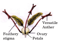
Given below is a flower with its characteristic features specialised for the
most common type of abiotic pollination.
Answer the following questions based on the above diagram :
(a). Name the mode of abiotic pollination that will be adopted by the given plant species in the above picture.
View Solution
Step 1: Understanding the Question:
The question asks to identify the mode of abiotic pollination based on the features of the flower shown in the diagram.
Step 3: Detailed Explanation:
The flower in the diagram displays several features characteristic of wind pollination:
- A large, feathery stigma.
- Well-exposed anthers (versatile).
- Small, inconspicuous petals.
These adaptations are designed to facilitate pollination by a non-living (abiotic) agent, specifically the wind. The technical term for wind pollination is Anemophily. Quick Tip: "Anemos" is Greek for wind. Anemophily literally means "wind-loving." An anemometer is a device that measures wind speed.
Describe the utility of the long, feathery, exposed stigma.
View Solution
Step 1: Understanding the Question:
The question asks for the functional advantage of the specific type of stigma shown in the diagram of a wind-pollinated flower.
Step 3: Detailed Explanation:
In wind pollination, the dispersal of pollen is non-directional and chancy. Therefore, the receptive organ (stigma) must be adapted to maximize the chances of capturing this airborne pollen.
- Feathery structure: This greatly increases the surface area of the stigma. It acts like a net or a sieve to filter pollen grains from the air that passes by.
- Long and Exposed: The stigma protrudes out of the flower to be in the direct path of wind currents, ensuring it is not shielded by other floral parts. Quick Tip: For wind pollination, the stigma is like a large fishing net—the bigger and more exposed it is, the more fish (pollen) it can catch from the vast ocean (air).
What two specific adaptations should the pollen grains have for this type of pollination?
1. Lightweight and non-sticky: To be easily carried by wind.
2. Produced in large quantities: To compensate for the high degree of wastage.
View Solution
Step 1: Understanding the Question:
The question asks for two adaptations of pollen grains that make them suitable for wind dispersal.
Step 3: Detailed Explanation:
1. Physical Properties: To travel long distances on air currents, the pollen must be very light and dry (non-sticky). Stickiness or heaviness would cause them to clump together and fall quickly, defeating the purpose of wind dispersal. Some, like pine pollen, even have wing-like structures to aid buoyancy.
2. Quantitative Production: Wind pollination is highly inefficient. The probability of any single pollen grain landing on a compatible stigma is extremely low. To counteract this, wind-pollinated plants produce an enormous number of pollen grains. This "safety in numbers" strategy ensures that, by chance, some pollen will successfully pollinate a flower. Quick Tip: Wind-pollinated pollen is like dust—light, dry, and produced in huge amounts to ensure some of it settles in the right place.
What could be the probable reason for the petals being small and inconspicuous (not brightly coloured)?
View Solution
Step 1: Understanding the Question:
The question asks why wind-pollinated flowers typically have small, dull petals.
Step 3: Detailed Explanation:
Features like large, brightly coloured petals, sweet nectar, and pleasant fragrances are all adaptations specifically evolved to attract biotic pollinators (animals). These features are signals and rewards for the animals who, in turn, transfer pollen.
Since wind is a non-living (abiotic) agent, it cannot be attracted by visual or chemical cues. For a wind-pollinated plant, producing large, showy petals would be a waste of energy and resources that could be better spent on producing massive amounts of pollen and large, effective stigmas. In fact, large petals could even be a hindrance by blocking wind flow to the stamens and stigma. Quick Tip: Form follows function in biology. If pollination is done by animals, the flower is an advertisement. If pollination is done by wind, the flower is a piece of functional aerodynamic equipment.
Explain the process of formation of placenta in a human female after the implantation of the blastocyst in the endometrium of the uterus.
View Solution
Step 1: Understanding the Question:
The question asks for a description of placentation—the process of forming the placenta—which occurs after the blastocyst has successfully implanted in the uterine wall.
Step 3: Detailed Explanation:
The formation of the placenta is a complex process involving both fetal and maternal tissues.
1. Post-Implantation Events:
- After the blastocyst embeds itself into the endometrium (the inner lining of the uterus), the outer layer of cells of the blastocyst, known as the trophoblast, begins to rapidly divide and expand.
2. Formation of Chorionic Villi:
- The trophoblast cells differentiate and develop into finger-like projections that invade deeper into the maternal endometrium. These projections are called the chorionic villi.
- Initially, these villi cover the entire surface of the blastocyst, but eventually, those on the side away from the uterine wall degenerate, while those embedded in the endometrium continue to grow and branch extensively. This forms the fetal portion of the placenta, known as the chorion.
3. Modification of Uterine Tissue:
- In response to the invading chorionic villi, the maternal uterine tissue at the site of implantation also undergoes significant changes. The endometrial cells, called decidual cells, enlarge and the maternal blood vessels erode.
- This erosion creates spaces or lacunae which fill with maternal blood. The chorionic villi become surrounded by and bathed in this pool of maternal blood.
4. Interdigitation and Placenta Formation:
- The chorionic villi (fetal tissue) and the uterine tissue (maternal tissue) become tightly interlocked or interdigitated.
- This intricate, composite structure formed by the intermixing of fetal and maternal tissues is the placenta. It establishes a very close connection between the two circulatory systems, although they do not directly mix. A thin membrane (the placental barrier) separates the fetal blood within the capillaries of the villi from the maternal blood in the surrounding spaces.
5. Function:
- This arrangement provides a large surface area for the efficient exchange of oxygen, nutrients, and hormones from mother to fetus, and carbon dioxide and metabolic wastes from fetus to mother.
Quick Tip: To remember the components of the placenta, think of it as a two-part structure:
- \textbf{Fetal Part:} Chorionic villi (originating from the trophoblast).
- \textbf{Maternal Part:} Uterine wall tissue (endometrium).
These two parts lock together like fingers ("interdigitation") to form the final organ.
Explain the biological treatment of primary effluent when passed into the large aeration tanks in a sewage treatment plant (STP).
View Solution
Step 1: Understanding the Question:
The question asks for an explanation of the biological process that takes place in the aeration tanks during secondary treatment at a Sewage Treatment Plant (STP).
Step 3: Detailed Explanation:
This stage of sewage treatment is known as Secondary Treatment or Biological Treatment. Its primary goal is to remove the dissolved organic matter from the sewage.
1. The Setup:
- The liquid portion from the primary settling tank, called the primary effluent, is pumped into large, open aeration tanks.
2. Aeration and Agitation:
- Inside these tanks, two key things happen:
a. Mechanical Agitation: The effluent is constantly stirred or agitated.
b. Aeration: Air is pumped into the tanks, providing a rich supply of oxygen.
3. Growth of Aerobic Microbes (Flocs):
- The combination of oxygen and a rich supply of organic matter (food) in the effluent creates ideal conditions for the rapid growth of aerobic bacteria.
- These bacteria associate with fungal filaments to form mesh-like, slimy structures called flocs. These flocs are the active biological component of this treatment phase.
4. Reduction of BOD:
- As the microbes in the flocs grow and multiply, they consume the dissolved organic waste present in the effluent.
- The amount of organic matter in water is measured by its Biochemical Oxygen Demand (BOD). BOD is the amount of oxygen that would be consumed if all the organic matter in one litre of water were oxidized by bacteria.
- By consuming the organic matter, the microbes significantly reduce the BOD of the sewage. A lower BOD indicates less pollution and cleaner water.
5. Settling of Flocs:
- Once the BOD has been reduced to a desired level (e.g., by 90-95%), the effluent is moved from the aeration tank to a settling tank.
- Here, the agitation and aeration are stopped. The bacterial flocs are allowed to settle down at the bottom as sediment. This sediment is called activated sludge.
- The clear liquid that remains on top, the secondary effluent, can then be discharged into natural water bodies or sent for tertiary treatment.
Quick Tip: Think of the aeration tank as a "microbial feast." You provide the microbes (bacteria in flocs) with two things they love: food (organic waste) and oxygen (aeration). In return, they "clean" the water for you by eating the waste, which is measured as a reduction in BOD.
Name the covalent bonds depicted as (a) and (b) in the form of slanting lines in the diagram.

View Solution
Step 1: Understanding the Question:
The question shows a simplified schematic of a DNA single strand and asks to identify two specific covalent bonds labeled as (a) and (b).
Step 3: Detailed Explanation:
In the structure of a polynucleotide chain:
- Bond (a): This bond connects the nitrogenous base (A, T, G) to the deoxyribose sugar (represented by the vertical line at the 1' carbon position). This specific covalent bond is known as an N-glycosidic bond.
- Bond (b): This bond links two adjacent sugar units together via a phosphate group, forming the sugar-phosphate backbone. It connects the 3' carbon of one sugar to the 5' carbon of the next sugar. This strong covalent linkage is called a phosphodiester bond.
Quick Tip: Remember, the strong \textbf{phosphodiester bonds} form the backbone of the DNA strand, giving it structural integrity. The \textbf{N-glycosidic bonds} attach the bases, which carry the genetic information, to this backbone.
How many purines are present in the given “stick” diagram ?
View Solution
Step 1: Understanding the Question:
The question asks to count the number of purine bases in the given DNA strand diagram.
Step 3: Detailed Explanation:
The nitrogenous bases in DNA are classified into two groups:
- Purines: Adenine (A) and Guanine (G).
- Pyrimidines: Cytosine (C) and Thymine (T).
The given diagram shows a DNA strand with the bases Adenine (A), Thymine (T), and Guanine (G).
- Adenine (A) is a purine.
- Guanine (G) is a purine.
- Thymine (T) is a pyrimidine.
Therefore, counting the purines, we find one Adenine and one Guanine, making a total of 2 purines.
Quick Tip: A simple mnemonic to remember the purines is "\textbf{Pur}e \textbf{A}s \textbf{G}old." This helps you recall that \textbf{Pur}ines are \textbf{A}denine and \textbf{G}uanine. The other two (in DNA) must be pyrimidines.
Draw the chemical structure of the given polynucleotide chain of DNA.
View Solution
Step 1: Understanding the Question:
The question asks to draw the detailed chemical structure of the DNA strand shown in the diagram, which has the sequence 5'-ATG-3'.
Step 3: Detailed Description of the Structure:
A chemical drawing would illustrate the following features:
Three Nucleotides: The chain consists of three nucleotide units.
Sugar-Phosphate Backbone: The backbone is formed by deoxyribose sugar molecules linked by phosphate groups. A phosphodiester bond connects the 5' carbon of one sugar to the 3' carbon of the previous sugar.
Nitrogenous Bases:
The first nucleotide (at the 5' end) has the base Adenine (A) attached to the 1' carbon of its sugar.
The second nucleotide has the base Thymine (T) attached to its sugar.
The third nucleotide (at the 3' end) has the base Guanine (G) attached to its sugar.
Polarity: The chain has a distinct 5' to 3' polarity.
The 5' end is marked by a free phosphate group attached to the 5' carbon of the first deoxyribose sugar.
The 3' end is marked by a free hydroxyl (-OH) group attached to the 3' carbon of the last deoxyribose sugar. Quick Tip: When drawing a polynucleotide chain, always start with the sugar-phosphate backbone. Then, attach the correct bases to the 1' carbon of each sugar. Finally, ensure you correctly label the 5' phosphate end and the 3' hydroxyl end to show the polarity.
Read the following passage and answer the questions that follow.The most convincing evidence to trace evolutionary relationships between humans and different groups of animals come from the basic similarities seen at the molecular level. Study the table given below that
depicts the number of amino acid differences between the haemoglobin polypeptide of few animals with that of humans and answer the questions that follow.

(a). To which category of evolution (Divergent or Convergent) do the following evolutionary relationships belong to :
(i) Humans and Macaque
(ii) Humans and Frog
View Solution
Step 1: Understanding the Question:
The question asks to classify the evolutionary relationship between humans and two other animals (macaque, frog) as either divergent or convergent, based on the provided context of molecular similarities in hemoglobin.
Step 3: Detailed Explanation:
- Divergent evolution occurs when two species sharing a common ancestor evolve and accumulate differences, resulting in the formation of new species. The underlying structures (like bones or, in this case, the hemoglobin molecule) are homologous (derived from a common ancestor) but may be modified for different functions.
- Convergent evolution is the independent evolution of similar features in species of different lineages. The structures are analogous, not homologous.
The table shows differences in hemoglobin, a molecule present in all these vertebrates, implying they share a common ancestor that had hemoglobin. The different numbers of amino acid changes reflect the degree of divergence from this common ancestry.
- (i) Humans and Macaque: Both are primates and share a relatively recent common ancestor. Their evolution from that ancestor is a clear case of divergence. The small number of amino acid differences (8) supports this close relationship.
- (ii) Humans and Frog: Both are vertebrates and share a much more distant common ancestor. They have diverged significantly over millions of years. The larger number of amino acid differences (67) reflects this greater evolutionary distance.
Since both pairs of organisms share a common ancestor and have accumulated differences over time, their relationship is a result of divergent evolution. Quick Tip: When comparing homologous structures (like the same protein in different species), the relationship is almost always one of divergent evolution. The degree of difference in the structure indicates how long ago the species diverged from their common ancestor.
What do the biochemical similarities in haemoglobin suggest about the evolutionary relationship between humans, frog and lamprey?
View Solution
Step 1: Understanding the Question:
The question asks what the molecular data on hemoglobin implies about the evolutionary history connecting humans, frogs, and lampreys.
Step 3: Detailed Explanation:
The fact that all three organisms—humans (mammal), frogs (amphibian), and lampreys (jawless fish)—possess hemoglobin for oxygen transport is a powerful piece of evidence for common descent.
Common Ancestry: It strongly suggests that they all evolved from a common ancestor that already used this protein for a similar function. Hemoglobin is a homologous molecule across these species.
Degree of Relatedness: The table shows the number of amino acid differences compared to humans: Frog (67 differences) and Lamprey (125 differences). This quantitative data allows us to infer the branching pattern of evolution. Since humans have fewer differences with frogs than with lampreys, it suggests that the human-frog lineage diverged more recently than the lineage leading to lampreys. The lamprey is the most distantly related of the three. Quick Tip: Molecular evidence, like comparing protein or DNA sequences, is a cornerstone of modern evolutionary biology. The fundamental principle is simple: more similarities = more closely related (i.e., more recent common ancestor).
Which one of the two – lampreys' or macaques' evolution is more closely related to humans and why ?
View Solution
Step 1: Understanding the Question:
The question asks us to use the provided data table to determine whether a lamprey or a macaque is a closer evolutionary relative to humans and to justify the answer.
Step 3: Detailed Explanation:
The basis for determining evolutionary relationships from molecular data is that as species diverge from a common ancestor, their DNA and protein sequences accumulate mutations over time. The number of differences, therefore, acts as a "molecular clock"—fewer differences imply that less time has passed since divergence, meaning a more recent common ancestor.
Data for Macaque: The difference in hemoglobin amino acids between a macaque and a human is 8.
Data for Lamprey: The difference between a lamprey and a human is 125.
Since 8 is a much smaller number than 125, it indicates that the lineage leading to humans and macaques diverged much more recently than the lineage leading to humans and lampreys. Therefore, the macaque is far more closely related to humans. This aligns with our understanding of classification, as both humans and macaques are primates, while lampreys are very primitive vertebrates. Quick Tip: In molecular evolution questions, always look for the smallest number of differences (in DNA or amino acids). The smallest number always points to the closest relative.
OR
Question 29 (c) (ii):
Which one of the two frogs' or dogs' evolution is more closely related to humans and why?
View Solution
Step 1: Understanding the Question:
The question asks us to use the data table to determine whether a frog or a dog is a closer evolutionary relative to humans and to justify the choice.
Step 3: Detailed Explanation:
We apply the same principle of the "molecular clock": fewer molecular differences mean a closer evolutionary relationship. We need to compare the amino acid differences for the dog and the frog with respect to humans.
Data for Dog: The difference in hemoglobin amino acids between a dog and a human is 32.
Data for Frog: The difference between a frog and a human is 67.
Comparing the two values, 32 is less than 67. This indicates that the common ancestor of humans and dogs lived more recently than the common ancestor of humans and frogs. Therefore, the dog is more closely related to humans than the frog is. This is consistent with biological classification, as both dogs and humans are mammals, while frogs are amphibians. Quick Tip: Always base your answer directly on the data provided. Even if you know from general biology that dogs are closer relatives to humans than frogs are, your justification must come from the numbers given in the table.
Read the following passage and answer the questions that follow.
Deaths related to the use of drugs were estimated at about 5,00,000 in
2019, 17 5 percent more than in 2009. Liver diseases attributed to
Hepatitis B are a major cause of drug-related deaths, according to
UNODC, accounting for more than half of the total number of deaths
attributed to the use of drugs. Drug overdoses account for a quarter of
drug-related deaths.
Opioids contribute to account for the most severe drug-related harm,
including fatal overdoses, when used non-medically. At the global level,
two-third of direct drug-related deaths are due to opioids, and in some
sub-regions the proportion can be as high as three-quarters of such
deaths.
(a). Why are people taking opioids more prone to liver diseases attributed to Hepatitis B ?
View Solution
Step 1: Understanding the Question:
The question asks for the link between opioid use and an increased risk of Hepatitis B-related liver disease, based on the provided passage.
Step 3: Detailed Explanation:
The passage links drug misuse with liver diseases attributed to Hepatitis B. Hepatitis B is a viral infection that primarily affects the liver. A major mode of transmission for the Hepatitis B virus (HBV) is through contact with infected blood or other body fluids.
A common method of opioid abuse is intravenous (IV) injection.
Individuals who inject drugs often share needles, syringes, and other drug preparation equipment.
If one person in the group is infected with Hepatitis B, the virus can contaminate the shared equipment.
Subsequent users of that equipment are then directly inoculated with the virus into their bloodstream.
This efficient mode of transmission leads to a much higher incidence of Hepatitis B among intravenous drug users compared to the general population, which in turn makes them more prone to the chronic liver diseases (like cirrhosis and liver cancer) that Hepatitis B can cause. Quick Tip: When thinking about drug abuse and diseases, always consider the mode of administration. Injection drug use is a high-risk behavior for all blood-borne pathogens, including Hepatitis B, Hepatitis C, and HIV.
What is meant by direct drug-related disease ?
View Solution
Step 1: Understanding the Question:
The question asks for the definition of a "direct drug-related disease" as implied in the passage.
Step 3: Detailed Explanation:
The passage contrasts deaths from diseases like Hepatitis B (which are indirectly related to drug use via transmission) with "direct drug-related deaths." A direct drug-related death is a consequence of the physiological and toxicological effects of the substance on the body.
The most prominent example given in the passage is a fatal overdose.
For opioids, an overdose typically causes severe respiratory depression—the drug suppresses the brain's control over breathing until it stops, leading to death from lack of oxygen.
This is a direct pharmacological effect of the drug, making it a direct drug-related death, as opposed to an indirect death from an infection acquired through drug-use practices. Quick Tip: Differentiate between direct and indirect harm from drugs. \textbf{Direct harm} = what the drug itself does to your body (e.g., overdose). \textbf{Indirect harm} = negative consequences of the behaviors associated with drug use (e.g., contracting infections, accidents).
What is the scientific name of the plant from which the opioids are derived and from which part of the plant is it extracted ?
View Solution
Step 1: Understanding the Question:
The question asks for the botanical source of opioids, specifically the scientific name of the plant and the part from which the raw material is obtained.
Step 3: Detailed Explanation:
Plant Source: Opioids are a class of drugs that are naturally found in the opium poppy plant. The scientific name for this plant is Papaver somniferum.
Extraction: The source of the opioids is the milky white fluid, or latex, that oozes out when the unripe seed pod (also called the capsule or fruit) of the poppy is scored or cut. This latex is collected and dried to produce raw opium. Raw opium contains a mixture of alkaloids, including morphine and codeine, from which other opioids (like heroin) can be synthesized. Quick Tip: Remember the connection: \textbf{Opium comes from the \textbf{Opium Poppy} (Papaver somniferum). The drug is in the \textbf{latex} from the unripe \textbf{poppy pod}.
OR
Question 30 (c) (ii):
State two common warning signs of drug abuse among the youth.
View Solution
Step 1: Understanding the Question:
The question asks for two common indicators or "warning signs" that may suggest a young person is abusing drugs.
Step 3: Detailed Explanation:
Drug abuse often leads to significant changes in a person's life and behavior. Common warning signs that parents, teachers, or friends might notice in an adolescent include:
Changes in School or Work Performance: This is a very common sign. It can include a sudden drop in grades, skipping classes, loss of motivation, or getting into trouble at school.
Changes in Social Circles and Behavior: The individual may withdraw from long-time friends and family members, become isolated and secretive, or start hanging out with a completely new group of friends. They might show a loss of interest in activities that were once important to them.
Changes in Mood and Personality: Unexplained mood swings, irritability, anger, depression, or a general lack of energy can be indicators.
Physical Signs: These can include a decline in personal grooming or appearance, unusual tiredness or hyperactivity, bloodshot eyes, and changes in appetite leading to weight loss or gain.
Financial Issues: A sudden, unexplained need for money, or stealing money or valuables from home, can be a sign that they are funding a drug habit.
The question asks for two, so any two from the above list would be correct. For example, a drop in academic performance and social withdrawal. Quick Tip: When looking for signs of drug abuse, the key word is "change." Look for sudden, unexplained, and negative changes in an adolescent's behavior, performance, social life, or physical appearance.
Give the full form of ‘ELISA'. Write the principle on which ELISA test is based.
View Solution
Step 1: Understanding the Question:
The question has two parts: first, to provide the full name for the acronym ELISA, and second, to explain the fundamental principle behind how this diagnostic test works.
Step 3: Detailed Explanation:
Full Form of ELISA:
The acronym ELISA stands for Enzyme-Linked Immunosorbent Assay. Let's break down what this means:
- Enzyme-Linked: An enzyme is chemically linked to an antibody or antigen.
- Immuno-: The test is based on an immune reaction (antigen-antibody binding).
- Sorbent: The antigens or antibodies are adsorbed (stuck) onto a solid surface, usually a microplate well.
- Assay: It is a test or analysis.
Principle of ELISA:
The core principle of ELISA is the highly specific antigen-antibody interaction. Here's a typical workflow for detecting an infection (i.e., detecting antibodies against a pathogen in a patient's blood):
1. Coating: A known antigen from the pathogen (e.g., a protein from HIV) is immobilized onto the surface of a plastic microplate well.
2. Sample Addition: The patient's blood serum is added to the well. If the patient has been infected, their serum will contain antibodies that specifically recognize and bind to the immobilized antigen.
3. Washing: The well is washed to remove any unbound antibodies.
4. Addition of Secondary Antibody: A second antibody is added. This "secondary antibody" is designed to bind specifically to human antibodies (the patient's antibodies). Crucially, this secondary antibody is covalently linked to an enzyme (e.g., horseradish peroxidase).
5. Washing: The well is washed again to remove any unbound enzyme-linked secondary antibodies.
6. Substrate Addition: A colorless substrate for the enzyme is added. If the enzyme-linked secondary antibody is present (which it will be if the patient's antibodies were present), the enzyme will act on the substrate and convert it into a colored product.
7. Detection: The development of color indicates that the patient's serum contained the specific antibodies, meaning they are infected. The intensity of the color can be measured to quantify the amount of antibody. Quick Tip: Remember the "sandwich" analogy for ELISA. The antigen is the bottom slice of bread. The patient's antibody is the "filling" that sticks to it. The enzyme-linked secondary antibody is the top slice of bread that sticks to the filling. The final substrate reaction adds the "color" to the sandwich, making it visible.
Describe all the methods used to treat or cure Adenosine deaminase deficiency in humans.
View Solution
Step 1: Understanding the Question:
The question asks for a comprehensive description of all available treatment options for ADA deficiency.
Step 3: Detailed Explanation:
ADA deficiency is a genetic disorder where the gene for the enzyme adenosine deaminase is defective. This enzyme is crucial for the function of the immune system, particularly lymphocytes. Its absence leads to a severe form of SCID. The treatment approaches aim to either replace the missing enzyme or correct the underlying genetic defect.
Bone Marrow Transplantation (BMT):
Principle: This method aims to provide a permanent source of healthy, enzyme-producing cells.
Procedure: The patient's bone marrow, which contains the defective hematopoietic stem cells, is ablated using chemotherapy. Then, healthy bone marrow containing functional stem cells from a histocompatible (tissue-matched) donor (ideally a sibling) is transplanted into the patient.
Outcome: If successful, the donor stem cells engraft and repopulate the patient's marrow, producing a lifetime supply of functional lymphocytes with the correct ADA gene. This is considered a permanent cure, but it carries risks like graft-versus-host disease and requires a suitable donor.
Enzyme Replacement Therapy (ERT):
Principle: This method provides the patient with the functional enzyme they cannot produce.
Procedure: The ADA enzyme, often modified with polyethylene glycol (PEG) to increase its lifespan in the body, is administered to the patient through regular intravenous injections.
Outcome: This therapy can restore immune function, but it is not a cure. The patient is dependent on periodic, lifelong injections as the enzyme is eventually degraded.
Gene Therapy:
Principle: This method aims to introduce the correct copy of the ADA gene into the patient's own cells.
Procedure: Lymphocytes (T-cells) are isolated from the patient's blood. A normal, functional copy of the human ADA gene is inserted into a retroviral vector. This vector is then used to infect the patient's lymphocytes in vitro, transferring the functional gene into their genome. The genetically modified lymphocytes are then cultured to increase their numbers and infused back into the patient.
Outcome: These corrected cells can temporarily restore the patient's immune function. However, because these mature lymphocytes have a limited lifespan, the patient requires repeated infusions. Thus, this approach is also not a permanent cure. A permanent cure through gene therapy would require modifying the hematopoietic stem cells themselves. Quick Tip: To organize the ADA deficiency treatments, think of the hierarchy of solutions: - \textbf{Level 1 (Symptomatic)}: Enzyme Replacement (just provides the missing protein). - \textbf{Level 2 (Cellular)}: Gene Therapy on lymphocytes (fixes some cells, but they die off). - \textbf{Level 3 (Curative)}: Bone Marrow Transplant (replaces the factory that makes all the cells).
OR
Question 31 (b):
Explain the process of separation and isolation of DNA fragments by a typical gel electrophoresis.
View Solution
Step 1: Understanding the Question:
The question asks for a step-by-step explanation of how agarose gel electrophoresis is used to both separate DNA fragments and then isolate a specific fragment.
Step 3: Detailed Explanation:
Agarose gel electrophoresis is a fundamental technique in molecular biology for separating macromolecules, especially DNA, based on their size.
Part 1: Separation of DNA Fragments
Casting the Gel: A gel is made by dissolving agarose powder (a polysaccharide from seaweed) in a buffer solution and heating it. As it cools, it solidifies into a porous, gelatinous matrix. A "comb" is placed in the molten agarose to create small pits or wells at one end.
Setting up the Apparatus: The solidified gel is placed in an electrophoresis tank and submerged in the same buffer solution, which conducts electricity.
Loading the Sample: The DNA samples, which have been previously cut with restriction enzymes, are mixed with a dense loading dye (which helps the sample sink into the well and allows tracking of the electrophoresis progress). The mixture is then carefully pipetted into the wells. A DNA ladder (a mixture of DNA fragments of known sizes) is usually loaded in one lane for comparison.
Applying Electric Current: An electric field is applied across the gel. The end with the wells is placed near the negative electrode (cathode), and the other end is near the positive electrode (anode).
Migration and Separation: DNA molecules have a net negative charge due to the phosphate groups in their backbone. Therefore, when the current is turned on, the DNA fragments are repelled by the negative electrode and migrate through the gel towards the positive electrode. The agarose gel matrix acts as a molecular sieve. Smaller DNA fragments can navigate through the pores of the gel more easily and thus move faster and farther than larger DNA fragments. This difference in migration speed results in the separation of the DNA fragments into distinct bands based on their size.
Part 2: Visualization and Isolation
Staining and Visualization: Since DNA is invisible to the naked eye, the gel must be stained to see the separated bands. The most common stain is ethidium bromide, an intercalating agent. After electrophoresis, the gel is soaked in a solution of ethidium bromide. When the stained gel is exposed to ultraviolet (UV) light, the DNA bands fluoresce and become visible as bright orange bands.
Isolation (Elution): To isolate a specific fragment, the band corresponding to the desired size (as determined by comparison with the DNA ladder) is identified under UV light. Using a clean scalpel or razor blade, this piece of the gel containing the DNA band is physically excised (cut out). The DNA is then extracted and purified from the agarose slice. This process of extracting DNA from the gel is known as elution. The purified DNA fragment is then ready for use in further procedures like ligation or PCR. Quick Tip: Remember the basic principle: In gel electrophoresis, DNA "runs to red." Since DNA is negatively charged, it moves towards the positive (red) electrode. The gel acts like a dense forest, and the DNA fragments are like runners: the smallest runners get through the forest the fastest.
Describe the population growth curve applicable in a population of any species in nature that has limited resources at its disposal.
View Solution
Step 1: Understanding the Question:
The question asks to describe the population growth pattern that occurs in a realistic scenario where resources are not infinite. This refers to the logistic growth model.
Step 3: Detailed Explanation:
The population growth curve that describes a population in an environment with limited resources is called the Logistic Growth Curve. This model is more realistic than the exponential model because it incorporates environmental limitations. The growth pattern unfolds in distinct phases:
Phase 1: Lag Phase
When a population is introduced to a new environment or is at a very low density, the initial growth is slow. This is because the individuals need time to adapt, mature, and find mates. The population size is small, so even if the per capita growth rate is high, the absolute increase in numbers is minimal.
Phase 2: Log Phase (or Exponential Growth Phase)
During this phase, resources (food, space) are plentiful, and environmental resistance (like competition, predation, disease) is low. The birth rate is much higher than the death rate. This leads to a period of rapid, accelerating, near-exponential growth. The population size increases dramatically.
Phase 3: Stationary Phase (or Plateau Phase)
As the population continues to grow, it starts to consume a significant portion of the available resources. This leads to increased competition among individuals. Predation and disease may also increase with population density. This is known as environmental resistance. Consequently, the population growth rate begins to slow down. The curve starts to flatten as it approaches the maximum population size that the environment can sustainably support. This maximum limit is called the carrying capacity (K). When the population size (N) equals the carrying capacity (K), the growth rate becomes zero (birth rate = death rate). The population then enters the stationary phase, where its size tends to fluctuate around K. Quick Tip: Think of logistic growth like a party in a small room. At first, when only a few people are there (lag phase), things are slow. As more people arrive, the party gets exciting and crowded quickly (log phase). Eventually, the room gets full (reaches carrying capacity), and the number of people coming in is balanced by the number of people leaving, so the crowd size stays stable (stationary phase).
Give the equation of this growth curve.
View Solution
Step 1: Understanding the Question:
The question asks for the mathematical equation that describes the logistic growth curve.
Step 2: Key Formula or Approach:
The formula for logistic growth modifies the exponential growth equation (dN/dt = rN) by adding a term that accounts for environmental resistance as the population approaches its limit.
Step 3: Detailed Explanation:
The Verhulst-Pearl Logistic Growth equation is given by: \[ \frac{dN}{dt} = rN \left( \frac{K-N}{K} \right) \]
Where:
\( \frac{dN}{dt} \) is the rate of change of population size (N) over time (t), i.e., the population growth rate.
\( r \) is the intrinsic rate of natural increase (the per capita growth rate under ideal, unlimited conditions).
\( N \) is the current population size.
\( K \) is the carrying capacity, the maximum population size the environment can support.
The term \( \left( \frac{K-N}{K} \right) \) represents the environmental resistance. When N is very small compared to K, this term is close to 1, and the growth is nearly exponential (rN). As N approaches K, this term approaches 0, causing the population growth rate to slow down and eventually stop. Quick Tip: Remember that the logistic equation is just the exponential equation (\(rN\)) multiplied by a "braking factor" (\( (K-N)/K \)). This factor represents the environmental resistance that slows growth as the population gets bigger.
Name the growth curve and depict a graphical plot for this type of population growth.
View Solution
Step 1: Understanding the Question:
The question asks for the name of the growth curve for a population with limited resources and a graphical representation of it.
Step 3: Detailed Explanation:
Name of the Curve:
This type of realistic population growth is called Logistic Growth. The curve it produces is known as a Logistic Curve, a Sigmoid Curve, or simply an S-shaped Curve due to its characteristic shape.
Graphical Plot:
A correct graphical plot should have the following features:
Axes: The x-axis is labeled "Time (t)" and the y-axis is labeled "Population Density (N)".
Carrying Capacity (K): A horizontal dashed or dotted line is drawn across the upper part of the graph and labeled "Carrying Capacity (K)". This line represents the maximum sustainable population.
The Curve: The plot of N versus t is an S-shaped curve.
It starts slowly near the x-axis (the lag phase).
It then becomes steeper as it rises, indicating rapid growth (the log or acceleration phase).
The curve's slope then decreases as it approaches the K line, indicating slowing growth (the deceleration phase).
Finally, the curve becomes horizontal and fluctuates around the K line (the stationary phase or plateau).
\begin{tikzpicture[scale=1.3, every node/.style={scale=0.8]
% Axes
\draw[->, thick] (0,0) -- (9,0) node[below left] {Time (t);
\draw[->, thick] (0,0) -- (0,5) node[left] {Population Density (N);
% Carrying Capacity Line
\draw[dashed, color=red, thick] (0,4) node[left] {K -- (9,4) node[right] {Carrying Capacity;
% The S-shaped curve
\draw[very thick, color=blue] (0.5,0.2) .. controls (2,0.5) and (4,3.5) .. (6,3.9) .. controls (7,4.1) and (8,4) .. (9,4);
% Label the curve
\node[blue, align=center] at (4,2) {Logistic Growth
(S-shaped Curve);
% Label phases
\node[align=center] at (2,0.9) {Lag
Phase;
\node[align=center] at (5,2) {Log
(Exponential)
Phase;
\node[align=center] at (7.5,3.5) {Stationary
Phase;
\end{tikzpicture Quick Tip: When asked to draw a growth curve, always label your axes (N vs. t) and clearly indicate the Carrying Capacity (K) with a dashed line. The shape is key: "J" for exponential, "S" for logistic.
OR
Question 32 (b) (i):
Explain the Species-Area relationship within a natural forest and also predict the nature of graph when species richness is plotted against the area for a wide variety of taxa.
View Solution
Step 1: Understanding the Question:
The question has two parts: explain the concept of the species-area relationship and predict the shape of its graph.
Step 3: Detailed Explanation:
Explanation of the Relationship:
The great German naturalist and geographer Alexander von Humboldt observed that the diversity of species is related to the size of the habitat. While exploring the South American jungles, he found that as he increased the area of his search, the number of different species he encountered also increased. This fundamental ecological pattern is known as the Species-Area Relationship.
The relationship is straightforward: larger areas can support more species than smaller areas. This is because larger areas tend to have:
Greater habitat diversity and more ecological niches.
Larger populations for each species, which reduces the risk of local extinction.
However, this increase is not linear. As you explore more and more area, the rate at which you find new species slows down. Eventually, you reach a point where expanding the area further adds very few, if any, new species.
Nature of the Graph:
When species richness (S) is plotted on the y-axis against the area (A) on the x-axis, the resulting graph for a wide range of organisms and geographical areas is consistently a rectangular hyperbola.
- Initial Steep Rise: For small areas, a small increase in area leads to a large increase in the number of species found.
- Flattening of the Curve: For larger areas, a large increase in area is needed to find even a few new species. The curve becomes less steep and approaches a plateau. Quick Tip: Think of searching for different types of candy in a house. If you start in one small drawer, you might find 3 types. Searching the whole room (larger area) might yield 10 types. Searching the entire house (even larger area) might yield 15 types. But searching the neighbor's house too might only add 2 more types. The rate of finding new types decreases as the search area gets bigger—this is the essence of the rectangular hyperbola in the species-area relationship.
Depict the graphical relationship between species richness and area.
View Solution
Step 1: Understanding the Question:
The question asks to depict the graph for the species-area relationship on a normal scale.
Step 3: Detailed Explanation of the Graph:
A correct graphical plot for the species-area relationship should have the following features:
Axes: The horizontal x-axis is labeled "Area (A)" and the vertical y-axis is labeled "Species Richness (S)".
The Curve: The graph is a curve that starts from the origin.
It rises sharply for low values of Area, showing that many new species are found when the explored area is small.
As the Area increases, the slope of the curve decreases. It continues to rise but becomes flatter and flatter. This shape is known as a rectangular hyperbola.
Equation on Graph (Optional but good practice): The curve can be labeled with its corresponding equation, S = cA\(^z\).
\begin{tikzpicture[scale=0.9, every node/.style={scale=0.9]
% Axes
\draw[->, thick] (0,0) -- (8,0) node[below left] {Area (A);
\draw[->, thick] (0,0) -- (0,5) node[left] {Species Richness (S);
% The rectangular hyperbola curve
\draw[very thick, color=blue] (0.5,0.5) .. controls (1,2) and (3,3.5) .. (7.5,4.2);
% Label the curve's equation
\node[blue, align=center] at (3.5,2.5) {S = cA\(^z\);
% Add a title
\node at (4, 4.7) {Species-Area Relationship;
\end{tikzpicture Quick Tip: When drawing the species-area graph, the key is the shape: it is \textbf{not} a straight line. It's a curve that shows diminishing returns. Make sure the curve is steep initially and then flattens out.
Give the equation of the Species-Area relationship for a wide variety of taxa on a logarithmic scale.
View Solution
Step 1: Understanding the Question:
The question asks for the mathematical equation of the species-area relationship when it is plotted on a log-log scale.
Step 2: Key Formula or Approach:
The standard equation for the species-area relationship is S = cA\(^z\). To find the equation on a logarithmic scale, we take the logarithm of both sides of this standard equation.
Step 3: Detailed Explanation:
The relationship between species richness (S) and area (A) is described by the equation: \[ S = cA^z \]
To transform this hyperbolic relationship into a linear one, ecologists use a logarithmic scale. By taking the logarithm of both sides of the equation, we get: \[ \log(S) = \log(cA^z) \]
Using the logarithmic rule \(\log(xy) = \log(x) + \log(y)\), we can expand this to: \[ \log S = \log c + \log(A^z) \]
Using the logarithmic rule \(\log(x^y) = y \log(x)\), we get the final linear equation: \[ \log S = \log c + z \log A \]
This equation is in the form of a straight line, \( y = mx + C \), where:
\( y = \log S \)
\( x = \log A \)
\( m = z \) (the slope of the line, also known as the regression coefficient)
\( C = \log c \) (the y-intercept)
This linear relationship on a log-log plot is extremely useful for ecological analysis and comparison between different regions or taxa. Quick Tip: Remember this key transformation: the species-area relationship is a \textbf{rectangular hyperbola} (\(S = cA^z\)) on a regular graph, but it becomes a \textbf{straight line} (\(\log S = \log c + z \log A\)) on a log-log graph. The slope of this line, 'z', is an important measure of biodiversity.
Explain how does double fertilisation take place in a flowering plant.
View Solution
Step 1: Understanding the Question:
The question asks for an explanation of the process of double fertilization, a hallmark of flowering plants (angiosperms).
Step 3: Detailed Explanation:
Double fertilization is a complex process that involves the fusion of male and female gametes, leading to the formation of both the embryo and its nutritive tissue.
Pollen Germination and Pollen Tube Growth: After a compatible pollen grain lands on the stigma, it absorbs moisture and germinates. It produces a thin tube, the pollen tube, which grows down through the style towards the ovary. The nucleus within the pollen grain divides to form two haploid (n) male gametes, which travel down the pollen tube.
Entry into the Ovule: The pollen tube enters the ovule, typically through an opening called the micropyle. It is guided by chemical signals from the synergid cells within the embryo sac.
Discharge of Male Gametes: The pollen tube penetrates one of the synergids and releases its two male gametes into the cytoplasm of the embryo sac.
The Two Fertilization Events:
First Fertilization (Syngamy): One of the two male gametes (n) moves towards the egg cell (n) and fuses with its nucleus. This fusion of the male and female gametes is true fertilization, or syngamy. It results in the formation of a diploid (2n) zygote.
Second Fertilization (Triple Fusion): The other male gamete (n) moves towards the large central cell of the embryo sac, which contains two haploid polar nuclei (n + n). This male gamete fuses with both polar nuclei. Since this event involves the fusion of three haploid nuclei (one from the male gamete and two polar nuclei), it is called triple fusion. This results in the formation of a triploid (3n) primary endosperm nucleus (PEN).
Because two distinct fusion events—syngamy and triple fusion—occur simultaneously within the embryo sac, this entire phenomenon is termed double fertilization. Quick Tip: To remember double fertilization, think "2 male gametes, 2 fusions": - Male Gamete 1 + Egg (n+n) \(\rightarrow\) Zygote (2n) \(\rightarrow\) Embryo. - Male Gamete 2 + 2 Polar Nuclei (n+n+n) \(\rightarrow\) Primary Endosperm Nucleus (3n) \(\rightarrow\) Endosperm (food).
Write the fate of the products of double fertilization in these plants.
View Solution
Step 1: Understanding the Question:
The question asks what the two structures formed during double fertilization—the zygote and the primary endosperm nucleus—develop into after the fertilization process is complete.
Step 3: Detailed Explanation:
The events of double fertilization lead to the formation of two key structures within the ovule, each with a specific developmental path (fate) that is crucial for the formation of a viable seed.
Fate of the Zygote (Diploid, 2n):
The zygote is the first cell of the new sporophytic generation. It is diploid and contains the combined genetic material from the male and female gametes. Following a period of dormancy, the zygote undergoes repeated mitotic cell divisions and differentiation (a process known as embryogenesis) to develop into the embryo. The embryo consists of a radicle (embryonic root), a plumule (embryonic shoot), and one or two cotyledons (seed leaves).
Fate of the Primary Endosperm Nucleus (Triploid, 3n):
The primary endosperm nucleus (PEN) is located within the central cell, which now becomes the Primary Endosperm Cell (PEC). The PEN divides repeatedly by mitosis to form a triploid nutritive tissue called the endosperm. The endosperm's primary function is to store food reserves (like starch, proteins, and oils). This stored food is used to provide nourishment to the growing embryo, either during its development within the seed or later during seed germination. In some plants (like cereals), the endosperm persists in the mature seed, while in others (like beans), it is completely consumed by the embryo before the seed matures.
Ultimately, the ovule containing the embryo and endosperm develops into the seed, and the surrounding ovary develops into the fruit. Quick Tip: A simple way to remember the fates: The \textbf{zygote} becomes the \textbf{baby plant (embryo)}, and the \textbf{endosperm} becomes the \textbf{baby's food (nutritive tissue)}.
OR
Question 33 (b) (i):
Explain the structure of testicular lobules in human male reproductive system. Name the two types of cells present in the seminiferous tubules and state their role.
View Solution
Step 1: Understanding the Question:
The question has two parts: first, to describe the anatomical structure of testicular lobules, and second, to identify the two main cell types within the seminiferous tubules and describe their functions.
Step 3: Detailed Explanation:
Structure of Testicular Lobules:
The human testis is an ovoid organ covered by a dense fibrous capsule called the tunica albuginea. Internally, this capsule extends septa (partitions) into the testis, dividing it into a number of compartments.
- These compartments are the testicular lobules.
- There are approximately 250 lobules in each testis.
- Within each of these lobules are located one to three highly coiled tubes known as the seminiferous tubules. These tubules are the actual sites of sperm production. The space between the seminiferous tubules is called the interstitial space, which contains blood vessels and Leydig cells (interstitial cells) that produce testosterone.
Cells of the Seminiferous Tubules:
The inner lining of each seminiferous tubule is composed of a specialized epithelium containing two main types of cells:
Male Germ Cells (Spermatogonia):
Identity: These are the diploid (2n) stem cells that give rise to sperm. They are located along the basement membrane on the periphery of the tubule.
Role: Their primary function is to undergo the process of spermatogenesis. They divide by mitosis to replenish their numbers and also differentiate into primary spermatocytes. These primary spermatocytes then undergo meiosis I and meiosis II to produce haploid (n) spermatids, which finally mature into spermatozoa (sperm).
Sertoli Cells (or Sustentacular Cells):
Identity: These are large, columnar, non-dividing cells that extend from the basement membrane to the lumen of the tubule, surrounding and embedding the developing germ cells.
Role: They are often called "nurse cells" because their main function is to support and nourish the developing sperm cells throughout spermatogenesis. They provide structural support, regulate the process, phagocytose excess cytoplasm shed by spermatids, and form the blood-testis barrier, which protects the germ cells from the immune system. Quick Tip: To remember the cells in the seminiferous tubule: - \textbf{Spermatogonia} = the "\textbf{G}enerators" of sperm. - \textbf{Sertoli} Cells = the "\textbf{S}upport" system for the developing sperm. Don't forget the cells outside the tubules in the interstitial space: Leydig cells, which produce testosterone.
Describe the role of hypothalamic hormone GnRH in spermatogenesis.
View Solution
Step 1: Understanding the Question:
The question asks for the specific role of the hypothalamic hormone GnRH in the process of sperm production (spermatogenesis).
Step 3: Detailed Explanation:
The process of spermatogenesis is under complex hormonal control, which is initiated and regulated by the hypothalamus. This control mechanism is often referred to as the Hypothalamic-Pituitary-Gonadal (HPG) axis.
GnRH Secretion: Spermatogenesis begins at puberty due to a significant increase in the secretion of a hormone called Gonadotropin-releasing hormone (GnRH) from the hypothalamus in the brain. GnRH is a peptide hormone that travels through a local portal blood system to its target organ.
Action on the Anterior Pituitary: The target for GnRH is the anterior lobe of the pituitary gland. The pulsatile release of GnRH stimulates the anterior pituitary to synthesize and release two crucial hormones known as gonadotropins.
Release of Gonadotropins (LH and FSH): The two gonadotropins released are:
Luteinizing Hormone (LH)
Follicle-Stimulating Hormone (FSH)
Action of LH and FSH on the Testes: These hormones then travel via the bloodstream to the testes to directly regulate spermatogenesis:
LH acts on the Leydig cells (interstitial cells) located in the spaces between the seminiferous tubules, stimulating them to synthesize and secrete androgens, primarily testosterone. Testosterone is the principal male sex hormone and is absolutely essential for stimulating and maintaining the process of spermatogenesis.
FSH acts on the Sertoli cells within the seminiferous tubules. It stimulates the Sertoli cells to secrete certain factors and Androgen-Binding Protein (ABP), which helps to concentrate testosterone within the tubules and aids in the process of spermiogenesis (the final stage of maturation where spermatids transform into motile spermatozoa).
In summary, GnRH does not act directly on the testes. Instead, it acts as the master regulator, initiating the entire hormonal cascade by stimulating the pituitary. Without GnRH, there would be no LH and FSH secretion, leading to a failure in testosterone production and Sertoli cell function, and thus, a failure of spermatogenesis. Quick Tip: Remember the chain of command for spermatogenesis:
1. \textbf{Hypothalamus} (The General) releases \textbf{GnRH}.
2. GnRH orders the \textbf{Anterior Pituitary} (The Officer) to release \textbf{LH} and \textbf{FSH}.
3. LH and FSH order the \textbf{Testes} (The Soldiers) to work:
- LH \(\rightarrow\) Leydig Cells \(\rightarrow\) Produce Testosterone.
- FSH \(\rightarrow\) Sertoli Cells \(\rightarrow\) Help sperm mature.
*The article might have information for the previous academic years, please refer the official website of the exam.